Kidney – structure and function
Kidney – structure and function. Biological principles in action. Learning Outcomes. 5.4.6 (a), (b) and (d). List main components of 3 body fluids Describe how to test for glucose, protein and urea Describe how to find concentration of urea in a solution

Kidney – structure and function
E N D
Presentation Transcript
Kidney – structure and function Biological principles in action
Learning Outcomes • 5.4.6 (a), (b) and (d). • List main components of 3 body fluids • Describe how to test for glucose, protein and urea • Describe how to find concentration of urea in a solution • Determine the urea concentration of a fluid • Outline the roles of the kidney in excretion and osmoregulation
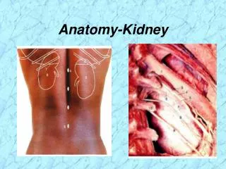
Kidney – structure and function • Where are they? • What are they for?
Roles of the kidney excretion homeostasis osmoregulation regulation of salts in the body regulation of pH production of a hormone (EPO)
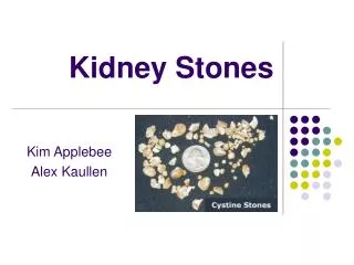
Testing Body fluids • You have three fluids labelled as X, Y and Z • You are provided with: • Clinistix / Diastix • Albustix • Urease and litmus paper • Find out what is in each of the three fluids.
Testing Body fluids • Draw out a flow chart to show how you would identify the following fluids using observations and simple laboratory tests like those you have just used: whole blood, plasma, serum, tissue fluid (filtrate), urine, bile, saliva.
Urea Determination Follow the instructions to produce a graph to determine the urea concentration of an unknown solution (U).
Urea Determination Answer questions (a), (b) and (c) and 8. Present as a coherent report. No need to reproduce the instructions, but you may if you wish.
Homework materials • Today’s work sheets • Homework Exercises • Useful Links Go to www.rfosbery-biology.co.uk Use: life, line, lifeline to enter the site Click on OHS, username is oxford, password is soapysam
Kidney dissection Learning outcomes • Describe the external features of the kidney • Describe the position of the kidneys in the body and relationships with blood supply and rest of u/g system • Draw and label LS kidney • Recognise different parts of the kidney • Make a drawing to scale
Kidney functions filtration of blood selective reabsorption by active transport passive absorption secretion
Kidney - structure Gross structure – what you can see with the naked eye Histology – what you can see through the microscope
Kidney – gross structure Position of kidneys in the body External structure Internal structure
Human kidney ureter renal artery renal vein attached here
Kidney – vertical section 1 = ureter 2 = pelvis 3 = cortex 4 = medulla
Histology of the kidney Learning outcomes • Find cortex, medulla and pelvis under the microscope • Describe the internal structure of the kidney • Draw a low power plan • Draw high power, labelled drawings of Mb, PCT, thick and thin loops, DCT and CD • Relate structure to function for the above • Make measurements with graticule eyepiece
Kidney – vertical section 1 = ureter 2 = pelvis 3 = cortex 4 = medulla
Kidney nephron cortex medulla name the parts?
branch of renal artery glomerulus Bowman’s capsule DCT PCT collecting duct branch of renal vein capillaries loop
Kidney – cortex (LP) glomerulus Bowman’s capsule proximal and distal convoluted tubules
Bowman’s capsule Glomerulus PCT
PCT microvilli DCT
Kidney - medulla • loops • collecting ducts • capillaries
Excretion and the kidneys Learning outcomes • State main excretory substances • Describe production and transport of urea • Explain why urea is produced • Explain why [salts] are regulated
Sources Where do these come from? • Water • Protein • Glucose • Urea • Uric acid • Creatinine • Ammonia
Sources • Water ingested drink and food / metabolic water • Protein ingested food / tissue breakdown • Glucose ingested food / glycogen / other compounds • Urea deamination / urea cycle • Uric acid metabolism of nucleotide bases • Creatinine metabolism of creatine (creatine phosphate) • Ammonia deamination
Urea formation • Excess protein / excess amino acids • Where from? • Deamination • Where? • Urea formation • Where? • Transport and excretion
Deamination • Oxidative deamination • Aerobic! • Liver (and other tissues) • Amino acid (glutamic acid) + oxygen • Keto acid + ammonia • Coupled with reduction of NAD (co-enzyme) • Ammonia!! Beware. • Ammonia enters the urea cycle • What happens to the keto acid?
Deamination Deamination is part of protein metabolism Catabolic reaction Details are at: http://www.elmhurst.edu/~chm/vchembook/632oxdeam.html
Urea/ornithine cycle • Ammonia comes from • deamination • and from aspartic acid produced from transamination • Carbon dioxide comes from link reaction and Krebs cycle • Urea is excreted • Requires ATP
Urea/ornithine cycle • Linked to: • deamination • transamination • Krebs cycle • phosphorylation of ADP (because ATP is required) • Details are at: http://www.elmhurst.edu/~chm/vchembook/633ureacycle.html
Protein metabolism • Deamination and urea cycle are part of the metabolism of proteins and amino acids in the body. More details of biochemistry (useful for MPB) at: http://www.elmhurst.edu/~chm/vchembook/index.html The link is on my web site for you.
Question 5 • Name? • Purpose? • Where? • Product • Intermediate (that gives its name to the cycle)
Sources Where do these come from? • Sodium • Potassium • Chloride • Phosphate • Sulphate
Sources Where do these come from? • Sodium extracellular cation • Potassium intracellular cation • Chloride extracellular anion • Phosphate bone / tissue fluid • Sulphate amino acids
Functions of the nephron Learning outcomes • Explain how ultrafiltration occurs relating structure to function • Explain how selective reabsorption occurs relating structure to function • Explain how structure of medulla is related to water potential gradients • Explain how water is reabsorbed throughout the nephron
Build a nephron • Sort the cards into three groups: • structures • substances • processes • Make a drawing/diagram of a nephron. • Use the structure cards to label it • Which ones are left over? • Use the substance cards to identify those carried into the kidney • Use the process cards to locate where these processes occur • You could use this approach to one of the tasks in your homework – BUT you don’t have to!
Processing in the kidneys • Ultrafiltration • Selective reabsorption • Secretion • Osmoregulation
Bowman’s capsule capillaries in the glomerulus
Ultrafiltration • blood pressure gives hydrostatic pressure that brings about filtration • capillaries have endothelium with pores • basement membrane is the filtration membrane • podocytes give support and do not provide resistance to filtration
lumen of Bowman’s capsule glomerulus
Ultrafiltration • Relate structure to function • Similar to filtration elsewhere in the body to produce tissue fluid • Composition of filtrate is similar to blood plasma. • What is missing?
Question 6 • X? • Y? • Z? Bullet points for (b) Explain…..
Kidney nephron cortex medulla
PCT microvilli DCT
Selective reabsorption • absorption of glucose, amino acids, ions, vitamins by PCT • absorption of ions by DCT • these are substances required by the body