Download
1 / 100
1.04k likes | 1.61k Views
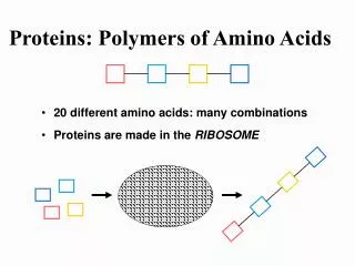
There is a huge number of different three-dimensional shapes possible, determined by the amino acid sequence of the polypeptide. Function follows structure. There is an enormous versatility in protein structure and therefore function.

E N D
There is a huge number of different three-dimensional shapes possible, determined by the amino acid sequence of the polypeptide. • Function follows structure. • There is an enormous versatility in protein structure and therefore function.
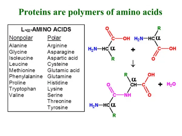
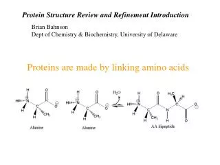
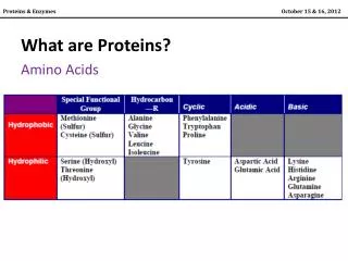
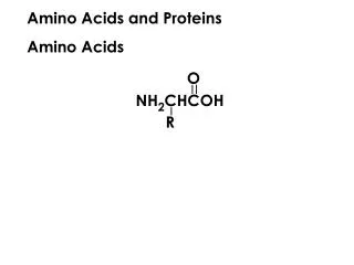
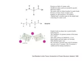
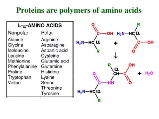
Proteins are made up of amino acids covalently bonded together by peptide bonds. alpha carbon amino terminus carboxyl terminus
Each protein has a unique sequence of amino acids. This amino acid sequence specifies the shape of the protein due to the fact that each protein folds into the most energetically favorable conformation
Many of the covalent bonds allow free rotation of the atoms they join. so that the polypeptide backbone can in principle fold up in an enormous number of ways. However each chain, depending on the sequence of amino acids will be constrained by many different sets of weak noncovalent bonds formed both by atoms in the polypeptide backbone and the atoms in the amino acid side chains. These weak bonds include hydrogen bonds, ionic bonds, van der Waals attractions. A fourth weak force important in protein folding is hydrophobic/hydrophilic interactions
The distribution of polar and nonpolar amino acids is important in how a protein folds. The nonpolar side chains tend to cluster in the interior of a molecule, avoiding contact with water, while the polar side chains arrange themselves near the outside.
Hydrophobic areas also tend to be found spanning the lipid bilayer of membranes like the plasma membrane.
enzyme lysozyme Large numbers of hydrogen bonds form between adjacent regions of the polypeptide chain and help stabilize its three-dimensional shape.
Each protein normally folds into a single stable conformation – of lowest energy. This conformation will change slightly during interactions with other molecules (as in enzyme-substrate complexes). This change in shape is often crucial for the function of the protein. Ex. receptor proteins See question 5-1 This conformation (the 3-D shape) is specified by its amino acid (aa) sequence. The non-covalent bonds and hydrophobic/philic interactions which hold a protein in the most energetically favorable conformation depend entirely on the aa sequence.
Molecular chaperones assists protein folding and prevent newly synthesized protein chains from associating with the wrong partners. Make protein folding more reliable. However, all the information required for proper protein folding is contained in its amino acid sequence. Proteins in a cell are found in a range of sizes. Protein sequencing has been replaced by DNA sequencing, which is much easier. Three-dimensional structure is determined by x-ray crystallography and NMR specroscopy. Panel 5-6, pg. 165.
C-terminus phosphocarrier protein HPr, a transport proteins that facilitates sugar transport into bacterial cells polypeptide backbone model N-terminus
Find two regular folding patterns: alpha helix and beta sheets. Both result from hydrogen-bonding between the N-H and C=O groups in polypeptide backbone, without involving side chains Every 4th peptide bond 1/2 bonds with 4-5 Complete turn every 3..6 aa Alpha helix is found in alpha-keratin, abundant in skin, hair, nails
The polypeptide chains are held together by hydrogen bonds between peptide bonds in different strands. The amino acid side chains in each strand alternately project above and below the plane of the sheet.
Antiparallel Parallel
Ex. Alpha-keratin, forms intracellulr fibers that reinforce the outer layer of the skin Coiled-coil And myosin molecules in muscle cells
Hydrophobic helices also tend to be found spanning the lipid bilayer of membranes like the plasma membrane. Channel proteins often have hydrophobic exteriors and hydrophilic interiors.
Domains are produced by any part of a polypeptide chain that can fold independently into a compact, stable structure - a modular unit. Proteins often have more than one domain - each with a specific function. Binds cyclic AMP (intracellular signaling molecule) Binds DNA Turns genes on or off Catabolite activator protein (CAP)
CAP is a bacterial signal transduction molecule • The large domain binds cyclic AMP, an intracellular signaling molecule. When cyclic AMP binds it causes a conformational change in the protein that enables the small domain to bind to a specific DNA sequence and turn on adjacent genes.
NAD-binding domain of the enzyme lactic dehydrogenase Cytochrome b, a single-domain protein involved in electron transfer in E. coli The variable domain of the immunoglobulin (antibody) light chain - a beta barrel Notice loops at each turn
The polypeptide chain generally passes back and forth across the entire domain, making sharp turns only at the protein surface. The protruding loop regions often form the binding sites for other molecules.
For each protein, a single conformation is extremely stable and has the exact chemical properties that enable the protein to perform a particular catalytic or structural function. • Proteins are so precisely built that the change of even a few atoms in one amino acid can sometimes disrupt the structure and the function of a protein. • Proteins can be grouped into families with very similar sequences and structures, probably due to genes duplicating and evolving.
Serine protease family: elastase, trypsin, chymotrypsin, and some proteases in blood clotting. Green portion: aa sequence is the same Notice the structural similarity and active site in red. Each cleaves different proteins or the bonds between different peptides Serine
Larger protein molecules may contain more than one polypeptide chain or subunit. The region that interacts with another molecule through noncovalent bonds is the binding site.
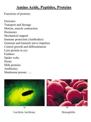
Hemoglbin contains two alpha globin subunits and two beta globin subunits. There are many large multi-subunit proteins in cells. Heme is the site where oxygen is carried
Proteins with one binding site can form a dimer Proteins with two different binding sites will often form a long helical filament or a closed ring
An actin filament, a helical array of actin proteins which can extent for micrometers in a cell - thousands of actin molecules Many large structures such as viruses and ribosomes are built from a mixture of different proteins plus RNA or DNA molecules. These structures can be isolated, dissociated, and often spontaneously reassemble into the original structure. Much of the structure of a cell is self-organizing.
Aminoacids - alpha helix Actin molecules - actin filaments Can be right or left handed (screw=right (clockwise) same when turned upsidedown
Ex. Alpha-keratin, a dimer, forms intracellular fibers (cytoskeleton) that reinforce the outer layer of the skin Coiled-coil Globular proteins fold up into compact shapes, like irregular ball. Most enzymes, even large and complex enzymes are globular. Fibrous proteins are elongated. Capped by globular domains which are binding sites, allowing assembly into ropelike, stable, intermediate filaments in skin, hair,horns.
Outside of the cell, fibrous proteins form the gel-like extracellular matrix. Secreted by cells, they assemble into sheets or long fibrils. Collagen, the most abundant in animal tissues, consists of three long polypeptide chains, each with the nonpolar aa glycine at every third position. Wind around each other in long regular triple helix and bind to one another side-by-side and end-to-end Elastin is formed from relatively loose polypeptide chains which are covalently cross-liked into a stretchy meshwork Skin, arteries lungs Hold tissues together.
Extracellular proteins are often stabilized by covalent cross-linkages. Esp disulfide bonds. These form as proteins are being exported from cells, catalyzed in the er by a special enzyme. Disulfide bonds do not typically form in the cell cytosol. They also do not change the protein’s conformation, but stabilize -reinforce - it.