Download

1 / 7
70 likes | 222 Views
Learn about the three categories of bacterial staining methods - simple, differential, and special stains - including the Gram stain technique and its step-by-step procedure. Understand the importance of staining to study bacterial morphology and cellular constituents.

E N D
Bacterial staining By Dr. Hesnaa Saeed Al Mossawi
STAINING • Stained preparations are needed in order to study their morphology and observe their cellular constituents • Smears can be made from liquid or solid cultures or from the clinical specimen • In Bacteriology, staining methods are divided into three categories • Simple stains: This makes use of the direct staining method.Differential stains: This staining method divides bacteria into two groupsSpecial stains: These are specialized staining methods to demonstrate certain bacterial components, e.g. spore.
Simple stain • Direct Staining: This a simple one-step staining procedure in which the presence and morphology of bacteria are demonstrated • Negative staining: This is when the organism remains unstained against a stained background. This is one of the few methods where acid stains such as nigrosin, are used
Gram stain SolutionsCrystal Violet: 0.5 to 1% in distilled water.Lugol's iodine: 10g Iodine 20g Potassium iodide1000ml Distilled waterDecolouriser:Absolute ethyl alcohol or acetone or acetone and alcohol mixture (1:1)CounterstainAqueous solution of neutral red or safranin 0.5%, or dilute carbolfuchsin. (1:10 dilution of strong carbolfuchin in distilled water)
Procedure • Make a smear, allow to dry and then fix with a gentle heat by passing the slide 2 or 3 times over a bunsen flame or placing the slide on a slide warmer.Stain with crystal violet for 1 minute.Wash with tap water.Apply Lugol's iodine and leave for 1 minute.Wash with tap water.Decolourisewith acetone or alcohol until no more colour appears to ooze out of the smear (about 1-2 seconds for acetone and 1-2 minutes for alcohol and 10 seconds for acetone/alcohol mixture) • Wash immediately with tap water. • Counterstain with neutral red or safranin or dilute carbolfuchsinfor 1 minute. • Wash with tap water. • Blot dry with a blotting or filter paper, and dry.
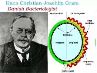
Gram stain • is the most important staining procedure. Gram positive bacteria stain purple, whereas gram-negative bacteria stain pink. • This difference is due to the ability of gram-positive bacteria to retain the crystal violet–iodine complex in the presence of a lipid solvent, usually acetone–alcohol. • Gram negative bacteria, because they have an outer lipid-containing membrane and thin peptidoglycan, lose the purple dye when treated with acetone–alcohol. • They become colorless and then stain pink when exposed to a red dye such as safranin