Download
1 / 71
710 likes | 788 Views
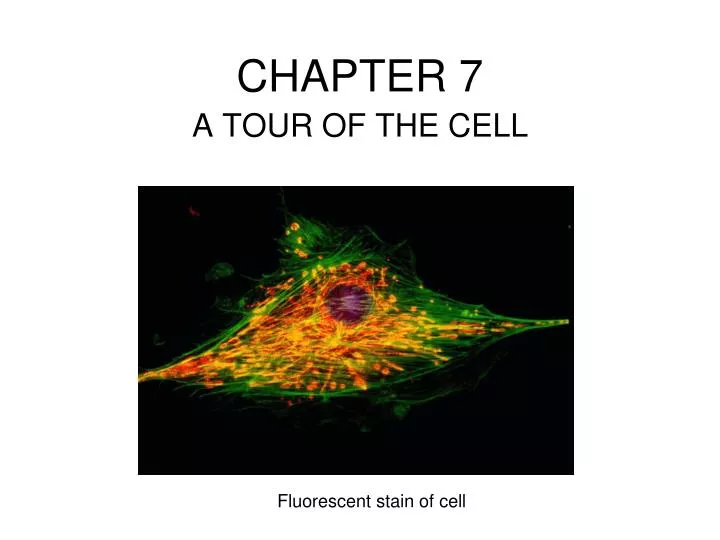
CHAPTER 7. A TOUR OF THE CELL. Fluorescent stain of cell. Cell Biology 1. The fundamental life processes of plants and animals depend on a variety of chemical reactions that occur in specialized areas of the organism’s cells. As a basis for understanding this concept, Students know:.

E N D
CHAPTER 7 A TOUR OF THE CELL Fluorescent stain of cell
Cell Biology1. The fundamental life processes of plants and animals depend on a variety of chemical reactions that occur in specialized areas of the organism’s cells. As a basis for understanding this concept, Students know: a. cells are enclosed within semipermeable membranes that regulate their interaction with their surroundings. b. enzymes are proteins that catalyze biochemical reactions without altering the reaction equilibrium and the activities of enzymes depend on the temperature, ionic conditions, and the pH of the surroundings. c. how prokaryotic cells, eukaryotic cells (including those from plants and animals), and viruses differ in complexity and general structure. d. the central dogma of molecular biology outlines the flow of information from transcription of ribonucleic acid (RNA) in the nucleus to translation of proteins on ribosomes in the cytoplasm. e. the role of the endoplasmic reticulum and Golgi apparatus in the secretion of proteins. f. usable energy is captured from sunlight by chloroplasts and is stored through the synthesis of sugar from carbon dioxide. g. the role of the mitochondria in making stored chemical-bond energy available to cells by completing the breakdown of glucose to carbon dioxide. h. Students know most macromolecules (polysaccharides, nucleic acids, proteins, lipids) in cells and organisms are synthesized from a small collection of simple precursors. i.* how chemiosmotic gradients in the mitochondria and chloroplast store energy for ATP production. j* Students know how eukaryotic cells are given shape and internal organization by a cytoskeleton or cell wall or both.
Organisms must exchange matter with the environment to grow, reproduce and maintain organization. Growth, reproduction and maintenance of the organization of living systems require free energy and matter. Molecules and atoms from the environment are necessary to build new molecules. • 1. Carbon moves from the environment to organisms where it is used to build carbohydrates, proteins, lipids or nucleic acids. Carbon is used in storage compounds and cell formation in all organisms. • 2. Nitrogen moves from the environment to organisms where it is used in building proteins and nucleic acids. • 3. Phosphorus moves from the environment to organisms where it is used in nucleic acids and certain lipids.
why are cells microscopic in size? • http://www.youtube.com/watch?v=xuG4ZZ1GbzI
The smaller the object, the greater Its ratio of surface area to volume. Metabolic requirements depend on passage of oxygen, nutrients and Carbon dioxide & other metabolic Waste through the plasma membrane. Geometric relationships explain why most cells are microscopic
why are cells microscopic in size? • Cell size is limited by the surface to volume ratio. • As cells get larger the volume increases at a greater rate compared to surface area. • Large cells can not get enough materials inside to stay alive.
b. Surface area-to-volume ratios affect a biological system’s ability to obtain necessary resources or eliminate waste products. • 1. As cells increase in volume, the relative surface area decreases and demand for material resources increases; more cellular structures are necessary to adequately exchange materials and energy with the environment. These limitations restrict cell size. • Ex. root hairs, cells of the alveoli, cells of the villi 2. The surface area of the plasma membrane must be large enough to adequately exchange materials; smaller cells have a more favorable surface area-to-volume ratio for exchange of materials with the environment.
villi cells within the small intestine • root hair cells of plants • cells of the alveoli within lungs • All shaped and arranged in ways that increase surface area to volume ratio and maximize diffusion.
what types of microscopes are used to view cells? • Light (2,000x) • Transmission Electron TEM (2,000,000x) • Scanning Electron SEM (3-D)
Rabbit trachea (windpipe) cell Transmission electron microscope (SEM) (TEM) Scanning electron microscope creates 3-D image of the surface of the same cell.
1665 1st Microscope • Robert Hooke discovered cells-cork • 1950’s Electron Microscope • Revealed the geography of the cell • ORGANELLES • Subcellular structures specialized • for various specific functions. • “tiny organ” • compartments or “rooms” • each contains specific enzymes
THINGS IN COMMON DIFFERENCES What is the difference between prokaryotic and eukaryotic cells?
Overview of a prokaryotic cell Overview of a plant cell Overview of an eukaryotic animal cell
Both have: Plasma membrane Cytosol- semifluid substance in which organelles are found. Chromosomes/genes Ribosomes (tiny organelles that make proteins according to instructions from the genes) Only eukaryotic cells: Have chromosomes inside a membrane bound organelle- the nucleus. “eu” = true “karyon” = kernel Are “large” -10x bigger than bacteria. Have other membrane-bound organelles. Prokaryotic Vs. Eukaryotic
FYI: CELL FRACTIONATION Technique used to determine the function of organelles. ORGANELLES are sub cellular structures that perform specific sets of chemical reactions for the cell within Eukaryotic Cells.
Eukaryotic cells maintain internal membranes that partition the cell into specialized regions. • a. Internal membranes facilitate cellular processes by minimizing competing interactions and by increasing surface area where reactions can occur. • b. Membranes and membrane-bound organelles in eukaryotic cells localize (compartmentalize) intracellular metabolic processes and specific enzymatic reactions. For example: • Endoplasmic Reticulum • Chloroplasts / Mitochondrion • Golgi • Nuclear envelope
CHARACTERISTICS • OF THE NUCLEUS: • Contains most of the genes* • Most conspicuous (big) part of the cell STRUCTURES of THE NUCLEUS (out to in): • Nuclear envelope (double membrane system- 2plbls) 2) Pores (protein tunnels) • Lamina (protein fiber scaffolding- network) 4) Chromatin (DNA & protein) 5) Nucleolus (makes ribosomes) *Chloroplasts and Mitochondria have their own DNA
Chromosomes are thick coiled chromatin fibers that condense when the cell is ready to divide. • Nucleosome = subunit of a chromosome… • DNA wrapped around 8 histone proteins.
Big Idea 4: Biological systems interact, and these systems and their interactions possess complex properties. The structure and function of subcellular components, and their interactions, provide essential cellular processes.
Figure 7.10 Ribosomes • RIBOSOMES are small universal structures (proks & euks) • made of ribosomal RNA (rRNA) and protein • carry out protein synthesis in 2 areas • free- suspended in the cytosol • bound- attached to the outside of the endoplasmic reticulum or nuclear envelope. • Ex. PANCREAS CELLS have a few million ribosomes • (synthesize: pancreatic juices, insulin, glucagon)
The Endoplasmic Reticulum • “within the cytoplasm” “little net” • Labyrinth of membrane tubes and sacs • > 1/2 the total membrane of the cell • Connected to the nucleus • FUNCTIONS: • Occurs in 2 forms: rough & smooth • rough ER provides site-specific protein synthesis with membrane-bound ribosomes • plays a role in intracellular transport = endomembrane system. • smooth ER synthesizes lipids.
Smooth ER Lacks ribosomes Functions: Synthesis of lipids sex hormones, oils, phospholipids 2) Metabolism of carbohydrates Detoxification of drugs/poisons LIVER CELLS MUSCLE CELLS (store Ca+) Rough ER Ribosomes attached to the cytoplasmic surface Functions: Protein synthesis on ribosomes, protein enters cisternal space to fold into native conformation. Secretory Glycoproteins Phospholipid membrane production (factory) Smooth ER vs. Rough ER ✘ Specific functions of smooth ER in specialized cells are beyond the scope of the course and the AP Exam.
Golgi ComplexAKA: golgi apparatus, golgi bodies • STRUCTURE membrane-bound, consists of a series of flattened membrane sacs (cisternae). • Looks like a smaller version of the ER but totally separate from nucleus) • FUNCTIONS include synthesis and packaging of materials (small molecules) for transport & production of lysosomes. • Receives & ships via transport vesicles- bags of membrane.
Golgi sorts, modifies, and exports cis “receiving” trans “shipping”
Lysosomes are membrane-enclosed sacs that contain hydrolytic enzymes, which are important in • intracellular digestion • The recycling of a cell’s organic materials and • programmed cell death (apoptosis) The formation and functions of lysosomes
lysosomes • Made by rough ER, finished in the Golgi • Contain hydrolytic enzymes that function at low pH • Pumps hydrogen ions from cytosol into lysosome to maintain acidic pH • Targets of primary lysosomes are: 1) food vacuoles (formed via phagocytosis) ex. Amoeba (protist) & Macrophages (white blood cells) 2) organelles or cytosol (autophagy- recycle materials) 3) Apoptosis = programmed destruction of cells ex. tadpole tail, human hand development- webbing • EX. Tay-Sachs genetic disorder is caused by missing/inactive lipid digesting enzyme which results in lipid accumulation in brain cells.
Review: relationships among organelles of the endomembrane system Endomembrane System Organelles that share membrane Components with each other. Nuclear Envelope, ER, Golgi, Lysosome, Vacuoles, and Plasma Membrane How? Transport Vesicles- little bag of Membrane.
Endomembrane System Rough ER vesicle Golgi Apparatus vesicle Plasma Membrane
The Golgi apparatus… stack of pita bread… insides = cisternae
VACUOLES • Larger than vesicles • Food vacuoles (formed by phagocytosis) • Contractile vacuoles- pump excess water out of the cell (freshwater protists) • Central vacuole- large vacuole in mature plant cells (membrane = tonoplast) - contains reserves of important compounds ie. pigments (petals), metabolic by-products (waste), poisons (repel predators), water, proteins and lipids (seeds)
Which cells have the larger vacuoles- animal or plant? The plant cell vacuole plant cells
Mitochondrion / mitochondria (pl) • Energy conversion organelle • Site of cellular respiration (x,y,z-->ATP) • Mitochondrial membrane proteins made by free ribosomes in the cytosol • Contain ribosomes and own DNA • Double membrane system - outer membrane smooth - inner membrane convoluted (cristae=folds) w/ proteins… increases surface area for rxns. • Two spaces: 1) mitochondrial matrix (inner most area) 2) inter membrane space- between the two membranes.
PLASTIDS • Family of closely related plant organelles. • Four kinds: • Chromoplasts- contain pigments that give fruits and vegetables their orange and yellow hues. • Leukoplasts- store starch, protein, oil • Amyloplasts- store starch (amylose) in roots and tubers. • Chloroplasts- contain green pigment chlorophyll & enzymes related to photosynthesis.
CHLOROPLAST STRUCTURE • Double membrane system • Pancakes in a “to-go” box • Thylakoids= flattened sacs (inside called “thylakoid space” • Grana= stacks of thylakoids • Stroma= area outside thylakoids and outer membrane… contains ribosomes, enzymes, and chloroplast DNA.
The chloroplast, site of photosynthesis * note: chloroplasts are larger than mitochondria.
PEROXISOMES • Specialized, one membrane, metabolic compartment that detoxifies substances. • Transfers hydrogen from substrates to oxygen- makes H2O2 • ie. detoxify alcohol or • use oxygen to break fatty acids into small molecules to be used as fuel for the mitochondria. • Contains catalase to convert H2O2 to water and Oxygen. • Liver cells have many.
The process of evolution drives the diversity and unity of life. Organisms are linked by lines of descent from common ancestry. Organisms share many conserved core processes and features that evolved and are widely distributed among organisms today. Structural evidence supports the relatedness of all eukaryotes. • Cytoskeleton (a network of structural proteins that facilitate cell movement, morphological integrity and organelle transport) • Membrane-bound organelles (mitochondria and/or chloroplasts) • Linear chromosomes • Endomembrane systems, including the nuclear envelope
THE CYTOSKELETON plays a major role in organizing the structures and activities of the cell made of: 1.microtubules 2.microfilaments 3.intermediate filaments
microtubules • structrure: • hollow fibers of tubulin = protein that makes microtubules - 2 types: alpha & beta (tubulin) • functions: 1) shape & support cell- compression resisting 2) tracks to move organelles equipped w/motor molecules. 3) assist in cell division (moving chromosomes) ex. Spindle fibers 4) motion for the cell- cilia/flagella
MICROTUBUELES grow out of : a centrosome (plant cells) 2 centrioles w/in the centrosome in (animal cells) centriole structure = 9 microtubule triplets in a ring
cilia & flagella:specialized microtubular structures • basal bodies (same structure as centrioles) anchor cilia and flagella to the cell membrane • flagella = long tails (few) 10-200 micrometers • cilia = short hairs (many) 2-20 micrometers (10-6) • structure of both… nine pairs of tubules arranged around 2 central tubules (“9 + 2”pattern in Euks) • Dynein = motor molecule (protein) attached to tubules, uses energy from ATP to move cilia. • Dynein “walking” = like a cat climbing a tree.
Figure 7.23 A comparison of the beating of flagella and cilia