Download
1 / 39
390 likes | 487 Views
Explore the world of proteins, from amino acids to complex shapes. Learn about protein functions, levels of structure, and denaturation. Additional resources and detailed information included.

E N D
Your Protein Structure Assignment 1. Define proteins and their function 2. What is an amino acid (monomers joined via dehydration synthesis) 3. How is a peptide bond formed? 4. What are the main uses of proteins in cells (plants and animals)??
Your Protein Shape Assignment 1. What are the various levels of protein ‘shape’? (primary, secondary, tertiary, quaternary) 2. How does structure relate to function with regard to proteins? 3. What does it mean to denature a protein?, Give one or more example.
Additional Resources (1) The Tree of Life, proteins and DNA module
Additional Resources (2) Protein structure and conformation links • Molecular Workbench DNA and protein module • http://www.youtube.com/watch?v=iaHHgEoa2c8&feature=related • http://www.youtube.com/watch?v=Q7dxi4ob2O4&feature=related
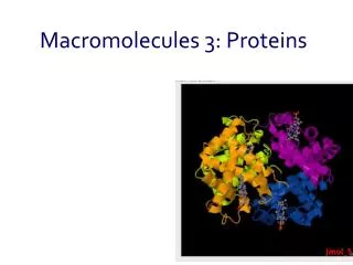
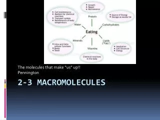
Proteins • > 50% of the dry mass of a cell is protein Proteins are used for: • Structural support • Energy storage • Transport of other substances • Signalling from one part of the organism to another • Movement • Defence against foreign substance • Enzymes • Humans have tens of thousands of different proteins • Most structurally sophisticated molecule, due to unique 3D shape or conformation
Amino Acid (Monomers) • Amino acid structure: NH3 - C - COOH • Amino acids differ due to the R (functional) group • The structure of the R-group determines the chemical properties of the amino acid
Proteins • Chemical composition C-H-O-N-(S) • Proteins are made up of smaller monomers called AMINO ACIDS • Amino Acids differ ONLY in the type of R (functional) group they carry Amino acids composed of 3 parts • Amino Group • Carboxylic group • Functional ®-group (Makes 20 different amino acids)
Hydrophilic Amino Acids Polar unchargedamino acids are hydrophilic & can form H-bonds • Serine • Threonine • Glutamine • Asparagine • Tyrosine • Cysteine
Hydrophobic Amino Acids Nonpolaramino acids are hydrophobic and are usually found in the center of the protein. They also found in proteins which are associated with cell membranes. • Glycine • Alanine • Valine • Leucine • Isoleucine • Methionine • Phenylalanine • Tryptophan • Proline)
Electrically charged Amino Acids The electrically charged amino acids have electrical properties that can change depending on the pH. • Aspartic Acid • Glutamic Acid • Lysine • Arginine • Histidine
Special Amino Acids • Cysteinecan form covalent disulfide bonds • Proline had a unique structure and causes kinks in the protein chain
Amino Acids link together to form polypeptides • 2 Amino Acids form a covalent bond, called a PEPTIDE BOND,through a condensation reaction to form a dipeptide • Multiple amino acids can bond to each other one at a time, forming a long chain called a POLYPEPTIDE
Protein shape • Each protein has a specific, and complex shape • Proteins are composed of one or more polypeptides • Different shapes allow proteins to perform different functions
Protein Shape Determines Function • Proteins with only primary and secondary structures are called fibrous proteins (claws, beaks, keratin, wool, collagen, ligaments, reptile scales) • Proteins with only 1,2,3 shapes are called globularproteins • If a protein is incorrectly folded, it can’t function correctly • Not understood how proteins fold themselves, seem to have molecules called chaperone proteins or chaperoninsthat assist others • A protein is denaturedwhen it loses its shape and therefore its ability to function correctly 20
Four Levels of Protein Structure/ Conformation 1. Primary- unique linear sequence in which amino acids are joined, can have dire circumstances if changed (insulin) 2. Secondary - refers to three dimensional shapes that are the result of H bonding at regular intervals, due to interactions between the amino acid backbones • alpha helix is a coiled shape • beta pleated sheet is an accordion shape 3. Tertiary Complex 3-D globular shape due to interactions between R groups of amino acids in it • Globular proteins such as enzymes are held in position by these interactions 4. Quaternary Consist of more than one polypeptide chain subunits, associated with interactions between these chains 19
Protein Conformation Primary Structure – sequence of amino acids Secondary structure – Folding and coiling due to H bond formation between carboxyl and amino groups of non-adjacent amino acid. R groups are NOT involved. Tertiary structure – disulfide bridges, ionic bonding, orH-bonding of R-groups Quaternary structure – 2+ amino acid chains R- group interactions, H bonds, ionic interactions
Primary Structure • A unique sequence of amino acids in a long polypeptide chain • Any changes in primary structure can affect a protein’s conformation and its ability to function • Example: Sickle cell anemia
Primary structure • The sequence of amino acids • Involves peptide bonds between the carboxyl and amine groups CYS LYS VAL PHE GLY ARG
Sickle cell anaemia • Sickling occurs due to a mutation of the Hb gene, associated with replacement of glutamic acid by valine
Secondary Structure • Segments of the polypeptide strand repeatedly coil or fold in a pattern which contributes to the overall conformation • Made by hydrogen bonds between the backbone of the amino acids (amino group and carboxyl groups) Structures formed include: • α-helices: area with a helical or spiral shape. Held together by H bonds between every 4th amino acid • β-pleated sheets:area where 2 or more regions of the polypeptide chain lie in parallel
Secondary structure • The amino acids in the primary structure can bond together to form : • a) An alpha helix b) a beta pleat • The bonds involved are hydrogen bonds • Large proteins will have regions containing both structures
Tertiary Structure Made of irregular contortions from interactions between side chains (R groups) • Hydrogen Bonds:between polar side groups • Ionic Bonds:between positively and negatively charged side chains • Hydrophobic Interactions:non-polar side chains end up on the inside of a protein, away from water—caused by water excluding these side chains from H bond interactions. Once together, held in place by dipole-dipole interactions • Disulfide Bridges:strong covalent bonds between cytosine’s sulfhydryl (-SH) groups
TERTIaRY STRUCTURE • The protein molecule undergoes further twisting and folding to form a 3 dimensional shape • The structure is held in place by interactions between R-groups of the different amino acids
Quaternary Structure The overall protein structure that results from the aggregation of 2 or more polypeptide subunits
QUATERNARY STRUCTURE • Proteins can contain more than one protein chain • E.g. immunoglobulins (form antibodies) • The bonds involved are the same as those for tertiary structure Chain 3 Chain 2 Chain 1
Denaturing of Protein Proteins can be denatured by: • Transfer from aqueous solution to an organic solvent (e.g. chloroform) • Any chemical that disruptsH-bonds, ionic bonds, & disulfide bridges • Excessive heat • Changes in pH
Denaturation • Protein conformation depends on the physical and chemical conditions of the protein’s environment • pH, salt concentration, temperature, and other aspects of the environment (aqueous or organic solvent) can unravel or change the conformation of the protein. • Change in protein shape causes it to lose its function • Some proteins can renatureand reform their conformation, other cannot.
TESTING FOR PROTEINS • Measure out 2cm3 of test solution into a test tube • Add 2 cm3 of Biuret solution • Shake and record colour change for each sample • Positive result = colour change from blue to lilac