Understanding the Diversity and Functions of Proteins: Fibrous and Globular Types
This overview explores the two main types of proteins: fibrous and globular. Fibrous proteins, such as collagen and keratin, have primary and secondary structures, are often water-insoluble, and provide structural support in tissues like tendons and skin. In contrast, globular proteins, like enzymes and hormones, possess all four levels of structure, are water-soluble, and perform essential functions including catalysis, transport, and regulation. Understanding protein structure and function is crucial for insights into biological processes and medical applications.

Understanding the Diversity and Functions of Proteins: Fibrous and Globular Types
E N D
Presentation Transcript
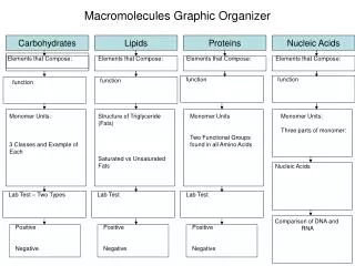
Fibrous (structural) proteins Only have primary and secondary structures • Water insoluble • VERY tough, may also be supple or stretchy • Parallel polypeptide chains in long sheets or fibres • STRUCTURAL proteins – collagen, cartilage, tendons, blood vessel walls • CONTRACTILE proteins – actin and myosin
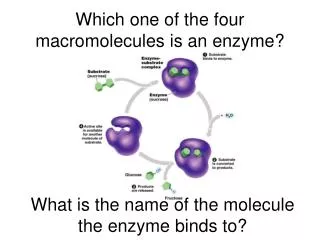
Globular proteins Have all four levels of protein structure • Water soluble • Tertiary structure critical to function • CATALYTIC (enzymes) • REGULATORY – hormones (insulin) • TRANSPORT (haemoglobin) • PROTECTIVE (immunoglobulins)
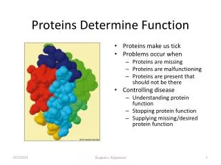
Proteins • > 50% of the dry mass of a cell is protein Proteins are used for: • Structural support • Energy storage • Transport of other substances • Signalling from one part of the organism to another • Movement • Defence against foreign substance • Enzymes • Humans have tens of thousands of different proteins • Most structurally sophisticated molecule, due to unique 3D shape or conformation
Types of protein • Structural support (Fibrous proteins) Silk: cocoons and webs Keratin: hair, horns, skin, nails, wool, beaks Collagen: tendons and ligaments
2.Globular proteins (e.g.Enzymes) Amylase Catalase Pepsin Trypsin DNA helicase DNA synthase Etcetc etc…
Globular proteins: Hormones • Insulin • ACTH • Vasopressin • Somatostatin • Prolactin • Growth hormone
Globular proteins:Transport proteins Haemoglobin, myoglobin: transport of essential substances (oxygen, carbon dioxide) Myoglobin was the first protein to be thoroughly described
Globular proteins: Energy storage Ovalbumin, Casein (milk protein), storage proteins in plant seeds
Movement proteins Actin and myosin form muscle fibres Animation of actin/myosin
Receptor proteins (also pumps, channel proteins) • Adrenergic receptors • G-protein receptors • Cannabinoid receptors • Opioid receptors • Aquaporin channels • Na/potassium pump proteins
8. Immune function:Antibodies (Immunoglobulins) Globular soluble proteins: IgG, gA, IgM,
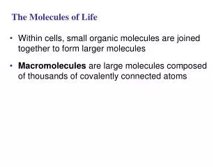
Amino Acid (Monomers) • Amino acid structure: NH3 - C - COOH • Amino acids differ due to the R (functional) group • The structure of the R-group determines the chemical properties of the amino acid
Proteins • Chemical composition C-H-O-N-(S) • Proteins are made up of smaller monomers called AMINO ACIDS • Amino Acids differ ONLY in the type of R (functional) group they carry Amino acids composed of 3 parts • Amino Group • Carboxylic group • Functional ®-group (Makes 20 different amino acids)
Amino Acids link together to form polypeptides • 2 Amino Acids form a covalent bond, called a PEPTIDE BOND,through a condensation reaction to form a dipeptide • Multiple amino acids can bond to each other one at a time, forming a long chain called a POLYPEPTIDE
Protein shape • Each protein has a specific, and complex shape • Proteins are composed of one or more polypeptides • Different shapes allow proteins to perform different functions
Protein Shape Determines Function • Proteins with only primary and secondary structures are called fibrous proteins (claws, beaks, keratin, wool, collagen, ligaments, reptile scales) • Proteins with only 1,2,3 shapes are called globularproteins • If a protein is incorrectly folded, it can’t function correctly • Not understood how proteins fold themselves, seem to have molecules called chaperone proteins or chaperoninsthat assist others • A protein is denaturedwhen it loses its shape and therefore its ability to function correctly 20
Four Levels of Protein Structure/ Conformation 1. Primary- unique linear sequence in which amino acids are joined, can have dire circumstances if changed (insulin) 2. Secondary - refers to three dimensional shapes that are the result of H bonding at regular intervals, due to interactions between the amino acid backbones • alpha helix is a coiled shape • beta pleated sheet is an accordion shape 3. Tertiary Complex 3-D globular shape due to interactions between R groups of amino acids in it • Globular proteins such as enzymes are held in position by these interactions 4. Quaternary Consist of more than one polypeptide chain subunits, associated with interactions between these chains 19
Primary Structure • A unique sequence of amino acids in a long polypeptide chain • Involves peptide bonds between the carboxyl and amine groups • Any changes in primary structure will affect a protein’s conformation and its ability to function • Example: Sickle cell anemia CYS LYS VAL PHE GLY ARG
Sickle cell anaemia Sickling occurs due to a mutation of the Hb gene, associated with replacement of glutamic acid by valine
Secondary Structure Made by hydrogen bonds between the backbone of the amino acids (amino group and carboxyl groups) • α-helices: area with a helical or spiral shape. Held together by H bonds between every 4th amino acid • β-pleated sheets:area where 2 or more regions of the polypeptide chain lie in parallel
αhelix a β-pleated sheet • The bonds involved are hydrogen bonds • Large proteins will have regions containing both structures
Tertiary Structure: FOLDING The protein folds up since various regions on the secondary structure are attracted to each other: • Disulfide Bridges:strong covalent bonds between cysteine’s sulfhydryl (-SH) groups • Ionic Bonds:between positively and negatively charged side chains • Hydrogen Bonds:between polar side groups • Hydrophobic Interactions:non-polar side chains end up on the inside of a protein, away from water
Quaternary Structure Complex proteins exist as aggregations of 2 or more polypeptide subunits
QUATERNARY STRUCTURE E.g. immunoglobulins • The bonds involved are the same as those for tertiary structure Chain 3 Chain 2 Chain 1
Protein denaturation Protein denaturation refers to loss of 3 – dimensional structure (and usually also biological function) of a protein – die to changing of the bonds that maintain secondary and 3rd degree structure, even though the amino acid sequence remains unaltered Denaturation can be caused by: • Strong acids and alkalis – profound pH change • Heavy metals – may disrupt ionic bonds • Heat, radiation, UV radiation • Detergents and solvents
Protein Conformation Primary Structure – sequence of amino acids Secondary structure – Folding and coiling due to H bond formation between carboxyl and amino groups of non-adjacent amino acid. R groups are NOT involved. Tertiary structure – disulfide bridges, ionic bonding, orH-bonding of R-groups Quaternary structure – 2+ amino acid chains R- group interactions, H bonds, ionic interactions
Primary Structure • A unique sequence of amino acids in a long polypeptide chain • Any changes in primary structure can affect a protein’s conformation and its ability to function • Example: Sickle cell anemia
Primary structure • The sequence of amino acids • Involves peptide bonds between the carboxyl and amine groups CYS LYS VAL PHE GLY ARG
Sickle cell anaemia • Sickling occurs due to a mutation of the Hb gene, associated with replacement of glutamic acid by valine
Secondary Structure • Segments of the polypeptide strand repeatedly coil or fold in a pattern which contributes to the overall conformation • Made by hydrogen bonds between the backbone of the amino acids (amino group and carboxyl groups) Structures formed include: • α-helices: area with a helical or spiral shape. Held together by H bonds between every 4th amino acid • β-pleated sheets:area where 2 or more regions of the polypeptide chain lie in parallel
Secondary structure • The amino acids in the primary structure can bond together to form : • a) An alpha helix b) a beta pleat • The bonds involved are hydrogen bonds • Large proteins will have regions containing both structures
Tertiary Structure Made of irregular contortions from interactions between side chains (R groups) • Hydrogen Bonds:between polar side groups • Ionic Bonds:between positively and negatively charged side chains • Hydrophobic Interactions:non-polar side chains end up on the inside of a protein, away from water—caused by water excluding these side chains from H bond interactions. Once together, held in place by dipole-dipole interactions • Disulfide Bridges:strong covalent bonds between cytosine’s sulfhydryl (-SH) groups
TERTIaRY STRUCTURE • The protein molecule undergoes further twisting and folding to form a 3 dimensional shape • The structure is held in place by interactions between R-groups of the different amino acids
Quaternary Structure The overall protein structure that results from the aggregation of 2 or more polypeptide subunits
QUATERNARY STRUCTURE • Proteins can contain more than one protein chain • E.g. immunoglobulins (form antibodies) • The bonds involved are the same as those for tertiary structure Chain 3 Chain 2 Chain 1
Denaturing of Protein Proteins can be denatured by: • Transfer from aqueous solution to an organic solvent (e.g. chloroform) • Any chemical that disruptsH-bonds, ionic bonds, & disulfide bridges • Excessive heat • Changes in pH
Denaturation • Protein conformation depends on the physical and chemical conditions of the protein’s environment • pH, salt concentration, temperature, and other aspects of the environment (aqueous or organic solvent) can unravel or change the conformation of the protein. • Change in protein shape causes it to lose its function • Some proteins can renatureand reform their conformation, other cannot.
TESTING FOR PROTEINS • Measure out 2cm3 of test solution into a test tube • Add 2 cm3 of Biuret solution • Shake and record colour change for each sample • Positive result = colour change from blue to lilac