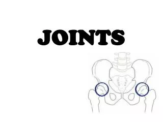
Joints
Chapter 8, Section 1. Joints. Joints, also called articulations , are functional junctions between two bones. The science of joints is called arthrology . Functions of joints Bind skeleton together Enable body movements Makes growth possible

Joints
E N D
Presentation Transcript
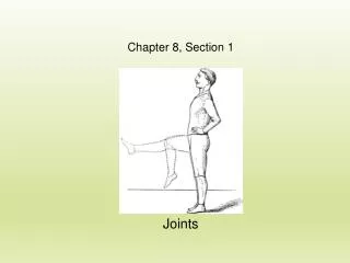
Chapter 8, Section 1 Joints
Joints, also called articulations, are functional junctions between two bones The science of joints is called arthrology. Functions of joints Bind skeleton together Enable body movements Makes growth possible Permit changes in skeleton for childbirth
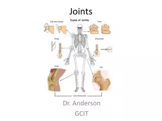
Classification of Joints Classifications based on amount of movement Synarthrotic = immovable Amphiarthrotic = slightly moveable Diarthrotic = fully movable Classifications by types of tissue: Fibrous joint = dense connective tissue Cartilaginous joint = bones connected by cartilage Synovial joint = contains a synovial membrane
Fibrous Joints • There are three (3) types of fibrous joints: • Syndesmosis • Suture • Gomphosis • 1. Syndesmosis: • Bones are connected by a sheet or bundle of fibrous tissue. • Examples include the interosseous membrane and interosseous ligaments between the tibia and fibula. Interosseous membrane between tibia and fibula is a syndesmosis joint.
Fibrous Joints • Suture: • Thin layer of dense • connective tissue • Connects flat bones of the • skull (sutural ligaments) • Synarthrotic • Gomphosis: • Cone-shaped bony process • in a bony socket • Example includes a tooth • anchored into a bony socket
Cartilaginous Joints • There are two (2) types of cartilaginous joints: • Synchondrosis • Symphysis • Synchondrosis • Bones are united by a band of hyaline cartilage. • Located between manubrium of sternum & 1st rib • Also located at epiphyseal plates of developing bone • Movement is synarthrotic
Cartilaginous Joints • Symphysis • A pad of fibrocartilage between two bones • Examples include the pubic symphysis and intervertebral discs • Movement is amphiarthrotic.
General Structure of Synovial Joints Synovial Joints are Freely movable (Diarthrotic) • Structures include: • Articular cartilage • Synovial membrane – secretes synovial fluid • Joint cavity – filled with synovial fluid • Joint capsule – dense connective tissue that stabilizes and protects joint Figure 8.7 The generalized structure of a synovial joint.
General Structure of Synovial Joints • Structures include: • Ligaments – bundles of collagenous fibers that reinforce the joint capsule • Menisci (sing. Meniscus) – pad of fibrocartilage that separates some joints. • Bursa – sac filled with synovial fluid. • Bursitis = inflammation of bursa Figure 8.8 Menisci separate the articulating surfaces of the femur and tibia. Several bursae are associated with the knee joint.
Types of Synovial Joints • Ball-and-socket • rounded head + cup-shaped socket. • Movement in all planes (multi-axial). • Allows for rotation • Includes hip joint and shoulder joint. • Condylar joint • Oval condyle + elliptical socket • Movements in most planes (bi-axial) • No rotational movement • Joints between metacarpals and phalanges/
Types of Synovial Joints • Plane (Gliding) Joint • Flattened bones slide across each other • Includes carpals and tarsals • ribs 2-7 articulate with sternum • Hinge joint • Increases or decreases angel between bones • Includes elbow joint • Joints between phalanges
Types of Synovial Joints • Pivot Joint • Rotation around a central axis (uni-axial) • Joint between radius and ulna • Joint between atlas (C1) and axis (C2). • Saddle Joint • 2 concave bones positioned at right angles • Includes metacarpal and carpal of thumb End of Chapter 8, Section 1
Section 2, Chapter 8 Types of Joint Movements
Types of Joint Movements Movement at a joint occurs when a muscle contracts and its fibers pull its moveable end (insertion) towards its fixed end (origin). Abduction = movement away from the midline (think of someone being abducted, or taken away) Adduction = movement towards the midline (think of adding together)
Types of Joint Movements • Flexion = decreases the angle of a joint • Bend elbow • Extension = increases the angle of a joint • Extend elbow • Hyperextension = extension beyond the anatomical position • bend hand back, bend head back beyond • anatomical position
Types of Joint Movements • Rotation = movement around a central axis • Twisting the head from side to side • Circumduction = movement so end follows a circular path • moving the finger in a circular motion • without moving the hand.
Types of Joint Movements • Elevation = raising a part • Shrugging the shoulders • Depression = lowering a part • Drooping the shoulders • Protraction = moving a part forward • thrusting head forward • Retraction = moving a part backward • pulling the head backward
Types of Joint Movements Supination = turning the hand so the palm faces upward or anteriorly Example: turning a doorknob clockwise with your right hand. Pronation = turning the hand so the palm faces downward or posteriorly Dorsiflexion = movement at the ankles that points toes towards the sky Plantar flexion = movement at the ankles that points toes towards the ground
Types of Joint Movements Eversion = turning the foot so the planter surface faces laterally Inversion = turning the foot so the plantar surface faces medially End of Chapter 8, Section 2
Chapter 8, Section 3 The Knee Joint and Joint Disorders
Knee Joint The knee joint is the largest and most complex synovial joint in body. Two distal condyles of the femur articulate with two proximal condyles of the tibia. This is a condylar joint. The femur also articulates anteriorly with the patella. This is a plane joint. Figure 8.21 Figure 8.20
Knee Joint General structures of a synovial joint in the knee • Synovial Membrane • Secretes synovial fluid • Joint Cavity • Stores synovial fluid • Joint Capsule • Relatively thin support • Reinforced by several ligaments and tendons Figure 8.20
Knee Joint Several ligaments and tendons strengthen the knee joint. Patellar tendon - The patella is partially enclosed in tendons fused together from the thigh muscle. Patellar ligament – continuation of patellar tendon. Extends from patella to the tibialtuberosity.
Knee Joint Ligaments continued: Tibial collateral ligament – connects medial condyle of femur with medial condyle of tibia. Fibular collateral ligament – connects lateral condyle of femur with head of fibula. Anterior & Posterior Cruciate ligaments – provide additional support to medial surface of tibia and femur Figure 8.21a Anterior right knee with patella removed.
Knee Joint Two menisci (medial & lateral meniscus) separate the femur and tibia, and align them. Figure 8.20 (a) sagittal section of the knee joint. (b) Photograph of the left knee joint (frontal section)
Knee Joint Three major bursae surround the knee joint. • Suprapatellar bursa • Largest bursa in body • Prepatellar bursa • Between patella and skin • Housemaid’s knee = prepatellar bursitis • Infrapatellar bursa Figure 8.20a
Joint Disorders Sprain = overstretching or tearing of connective tissue (tendons, ligaments, or cartilage) associated with a joint. However, the bones are not disarticulated.
Joint Disorders Arthritis = inflamed, swollen, and painful joints. • Osteoarthritis • Most common arthritis • Occurs with aging • Articular cartilage degenerates, causing bone to rub against bone. • Results in stiff and painful joints • Fingers may appear gnarled and knee may bulge. Osteoarthritic fingers often take on a gnarled appearance.
Joint Disorders • Rheumatoid Arthritis • Autoimmune disorder (immune system attacks tissue) • Synovial membrane thickens & becomes inflamed • Mass of fibrous connective tissue (Pannus) invades synovial space. • Fibrous pannus destroys articular cartilage, and the joints may swell and ossify. Knuckles may swell as a result of rheumatoid arthritis. • Other symptoms of Rheumatoid Arthritis: • low-grade fever, fatigue, appetite, stiffness. End of Chapter 8, Section 3