Joints
Joints. Hip region Knee region Ankle region. Hip region. sacroiliac joints. hip joint. pubic symphysis. Hip region. Hip region. hip joint. Knee region. Knee joint. Menisci Medial meniscus lateral meniscus


Joints
E N D
Presentation Transcript
Joints • Hip region • Knee region • Ankle region
Hip region sacroiliac joints hip joint pubic symphysis
Hip region hip joint
Knee region Knee joint • MenisciMedial meniscus lateral meniscus • Ligaments Cruciate ligaments Collateral ligaments Patellar ligament oblique popliteal ligament • Joint capsuleSynovial membrane Fibrous membrane
Knee region Minisci : fibrocartilaginous ‘shock absorbers’
Knee region Attachments
Knee region 1. The medial meniscus is more injured than the lateral meniscus, why?
Knee region Ligaments • anterior cruciate ligament (ACL) • posterior cruciate ligament (PCL) • medial collateral ligament (MCL, tibial collateral ligament) • lateral collateral ligament (LCL , fibular collateral ligament) • oblique popliteal ligament • ligamentum patellae
Knee region Cruciate ligaments ACL: Anteromedial part of the intercondylar area of tibia to inner aspect of lateralcondyle of femur. PCL: Posterolateral part of the intercondylar area of the tibia to inner aspect of medialcondyle of femur.
Knee region AnteriorCruciate ligament prevents anterior displacement of the tibia in relation to femur in flexion.
Knee region PosteriorCruciate ligament prevents posterior displacement of the tibia in relation to femur in extension.
Knee region How to test ACL and PCL? Drawer sign test
Knee region Anterior Drawer Test for ACL • Physician Position & Movements • Patient Position Note direction of forces
Knee region Posterior Drawer Testing- PCL Note direction of forces
Knee region Collateral ligaments
Knee region MCL attaches to the medial meniscus 2. The medial meniscus is more injured than the lateral meniscus, why?
Knee region Collateral ligaments action • Protect the knee joint from bending side to side. • Helps the locking mechanism
Knee region 3. The medial meniscus is more injured than the lateral meniscus, why?
Knee region Locking mechanism:less energy to maintain the standing position • Medial rotation of femur on the tibia during full extension tightencolateral ligaments.
Knee region Locking mechanism (2) • Joint surfaces become larger and more stable in extension.
Knee region Locking mechanism (3) • body's center of gravity is positioned along a vertical line that passes anterior to the knee joint.
Knee region Collateral ligaments test
Knee region Test for MCL Note Direction Of Forces
Knee region Test for LCL Note direction of forces
Knee region Patellar ligament Strengthening Anterior Aspect of Knee Joint
Knee region oblique popliteal ligament
Knee region Review • (1) patellar ligament • (2) tibial (medial) collateral ligament • (3) fibular (lateral ) collateral ligament • (4) medial meniscus • (5) lateral meniscus • (6) anterior cruciate ligament • (7) posterior cruciate ligament
Knee region Joint fibrous capsule
Knee region Joint synovial membrane
Knee region Bursa • little fluid sacs that helps the muscles and tendons slide freely:PrepatellarInfrapatellarSuprapatellar
Knee region Bursa
Knee region Prepatellar Bursitis
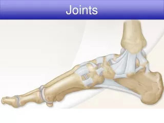
Ankle region • Ankle joint (talocrural joint) • Subtalarjoint (ST J.) • Talocalcaneonavicular joint (TCN J.)
The ankle, or talocrural joint • Needs ligaments!
The ankle, or talocrural joint, is a hinge joint • Plantar Flexion • Dorsi Flexion Inversion/Eversion?
Intertarsal joint: • Subtalar joint (ST J.) • Talocalcaneonavicular joint (TCN J.) • Calcaneocuboid (small rotation) • Naviculoconeiforms (almost no movement)