Synovial Joints Overview: Shoulder, Elbow, Wrist, Hip, Knee, Ankle
Explore the anatomy and function of various synovial joints from the shoulder to the ankle. Learn about common injuries and conditions affecting these joints.


Synovial Joints Overview: Shoulder, Elbow, Wrist, Hip, Knee, Ankle
E N D
Presentation Transcript
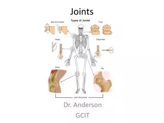
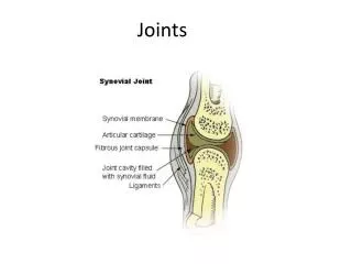
Part 5 Joints
Selected Synovial Joints • Shoulder (Glenohumeral) joint • The most freely movable joint lacks stability • Articular capsule is thin and loose • Muscle tendons contribute to joint stability
Glenohumeral Joint Figure 9.8a
Glenohumeral Joint • The rotator cuff is made up of four muscles and their associated tendons • Subscapularis • Supraspinatus • Infraspinatus • Teres minor • Rotator cuff injuries are common shoulder injuries
The shoulder joint Figure 9.8b, c
The Shoulder Joint Figure 9.8d, e
Selected Synovial Joints • Elbow joint • Allows flexion and extension • The humerus’ articulation with the trochlear notch of the ulna forms the hinge • Tendons of biceps and triceps brachii provide stability
Elbow Joint Figure 9.9a, b
Elbow Joint Figure 9.9c, d
Wrist Joint • Stabilized by numerous ligaments • Composed of radiocarpal and intercarpal joint • Radiocarpal joint – joint between the radius and proximal carpals (the scaphoid and lunate) • Allows for flexion, extension, adduction, abduction, and circumduction • Intercarpal joint – joint between the proximal and distal rows or carpals • Allows for gliding movement
Wrist Joint Figure 9.10a
Wrist Joint Figure 9.10b
Wrist Joint Figure 9.10c
Selected Synovial Joints • Hip joint • A ball-and-socket structure • Movements occur in all axes • Limited by ligaments and acetabulum • Head of femur articulates with acetabulum • Stability comes chiefly from acetabulum and capsular ligaments • Muscle tendons contribute somewhat to stability PLAY Movement at the hip joint: An overview
Frontal Section and Anterior View of the Hip Joint Figure 9.11a, b
Posterior View of the Hip Joint Figure 9.11c, d
Selected Synovial Joints • Knee joint • The largest and most complex joint • Primarily acts as a hinge joint • Has some capacity for rotation when leg is flexed • Structurally considered compound and bicondyloid • Two fibrocartilage menisci occur within the joint cavity
Sagittal Section and Superior View of Knee Joint Figure 9.12a
Sagittal Section and Superior View of Knee Joint Figure 9.12b
Knee Joint • Capsule of knee joint • Covers posterior and lateral aspects of the knee • Covers tibial and femoral condyles • Does not cover the anterior aspect of the knee • Anteriorly covered by three ligaments • Patellar, medial, and lateral retinacula
Anterior View of Knee Figure 9.12c
Knee Joint • Ligaments of the knee joint • Become taut when knee is extended • These extracapsular ligaments are • Fibular and tibial collateral ligament • Oblique popliteal ligament • Arcuate popliteal ligament
Posterior View of Knee Joint Figure 9.12d
Knee Joint • Intracapsular ligaments • Cruciate ligaments • Cross each other like an “X” • Each cruciate ligament runs from the proximal tibia to the distal femur • Anterior cruciate ligament • Posterior cruciate ligament
Anterior View of Flexed Knee Figure 9.12e, f
Knee Joint • Cruciate ligaments • Prevent undesirable movements at the knee joint Figure 9.13a
Selected Synovial Joint • Ankle Joint • A hinge joint between • United inferior ends of tibia and fibula • The talus of the foot • Allows the movements • Dorsiflexion and plantar flexion only
The Ankle Joint • Medially and laterally stabilized by ligaments • Medial (deltoid) ligament • Lateral ligament • Inferior ends of tibia and fibula are joined by ligaments • Anterior and posterior tibiofibular ligaments
The Ankle Joint Figure 9.15a
Ligaments of the Ankle Joint Figure 9.15b
Ligaments of the Ankle Joint Figure 9.15c
Ligaments of the Ankle Joint Figure 9.15d
Selected Synovial Joints • Temporomandibular joint (TMJ) • Lies anterior to the ear • Head of the mandible articulates with the mandibular fossa • Two surfaces of the articular disc allow two kinds of movement • Hinge-like movement • Superior surface of disc glides anteriorly
The Temporomandibular Joint Figure 9.16a, b
Selected Synovial Joints • Sternoclavicular Joint • Is a saddle joint • Muscles and ligaments contribute to joint stability • Unique joint shape allows for multiple complex movements • Another example of a saddle joint • Joint between trapezium and metacarpal 1
Sternoclavicular Joint Figure 9.17a
Sternoclavicular Joint Figure 9.17b
Disorders of Joints • Structure of joints makes them prone to traumatic stress • Function of joints makes them subject to friction and wear • Affected by inflammatory and degenerative processes
Joint Injuries • Sprains – ligaments of a reinforcing joint are stretched or torn • Dislocation – occurs when the bones of a joint are forced out of alignment • Torn cartilage – common injury to meniscus of knee joint
Inflammatory and Degenerative Conditions • Bursitis – inflammation of a bursa do to injury or friction • Tendonitis – inflammation of a tendon sheath
Inflammatory and Degenerative Conditions • Arthritis – describes over 100 kinds of joint-damaging diseases • Osteoarthritis – most common type “wear and tear” arthritis • Rheumatoid arthritis – a chronic inflammatory disorder • Gouty arthritis (gout) – uric acid build-up causes pain in joints • Lyme disease – inflammatory disease often resulting in joint pain
The Joints Throughout Life • Synovial joints develop from mesenchyme • By week 8 of fetal development, joints resemble adult joints • Outer region of mesenchyme becomes fibrous joint capsule • Inner region becomes the joint cavity
The Joints Throughout Life • During youth – injury may tear an epiphysis off a bone shaft • Advancing age – osteoarthritis becomes more common • Exercise – helps maintain joint health