Download
1 / 70
700 likes | 755 Views
The immune system is a specialized form of connective tissue distributed throughout the body, protecting against microorganism invasion. Discover its components like lymphocytes, thymus, and more. Explore primary and secondary organs, including the spleen and lymph nodes.

E N D
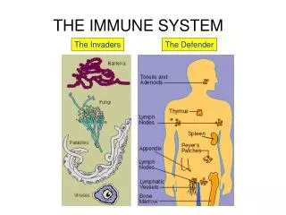
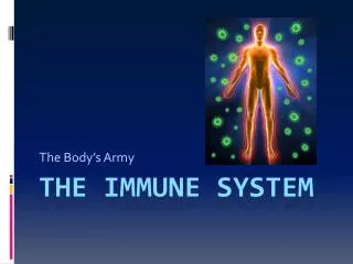
It is a specialized form of connective tissue that consists of groups of cells, tissues and organs distributed throughout the body.Their function is to protect the body from the invasion and damage by microorganisms and foreign materials by destroying or neutralizing them.
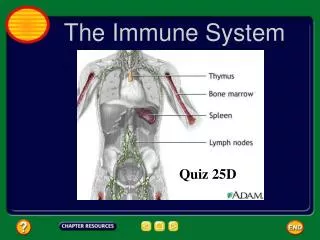
This system includes:Spleen ,thymus,lymph nodes,tonsils, Peyer’s patches, lymphatic(lymphoid) nodules and diffuse lymphatic tissue. • The cells, tissues and organs of the immune system are supported by reticular fibers(collagen III) formed by reticular cells.
The cells of the immune system : • Lymphocytes,macrophages, plasma cells, granulocytes and antigen presenting cells.These cells communicate with each other and with cells of other systems through signaling proteins called cytokines.
The cells of the immune have the ability to distinguish self ( the individuals own macromolecules) from nonself (foreign substances).
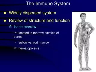
In early fetal life, the precursor cells which give rise to stem lymphoctes, originate in the yolk sac, liver and spleen.By late fetal life and the postnatal period the precursor cells are restricted to the bone marrow.
The undifferentiated stem lymphocyes migrate to sites in the bone marrow(B lymphocytes) or to the thymus(T lymphocytes) where the undergo differentiation into immunocompetent cells. Therefore the bone marrow and thymus are primary organs.
From the primary organ T&B lymphocytes then migrate to the rest of the lymphatic organs and tissues where they undergo antigen- dependent differentiation and proliferation into effector and memory. These organs are called secondary organs.
Classification: • 1. Primary(Central) e.g Thymus, Bone Marrow • Secondary(Peripheral) e.g Spleen , Lymph Nodes , Tonsils, Lymphatic nodules, Diffuse lymphatic tissues
2. Encapsulated or Unencapsulated. • The encapsulated organs are spleen, lymph nodes and thymus. The rest are unencapsulated.
The thymus is a primary lymphoid organ in that it supplies other lymphoid organs and tissues with T-lymphocytes. • The thymus is enclosed by a thin c.t.capsule from which numerous septa extend into the thymus subdividing the 2 lobes into numerous lobules.
Each lobule is divided into: • Cortex : darker peripheral zone because lymphocytes are more abundant in the cortex. • Medulla lighter central zone because epithelial reticular cells are more abundant in the medulla.
Epithelial reticular cells in the medulla form Hassal's corpuscles. Their cytoplasmic extensions form support for parenchyma and blood vessels forming the blood- thymus barrier. Macrophages are also present in addition to the lymphocytes and epithelial reticular cells.
Lymph Node It is an encapsulated secondary lymphoid organ. The capsule also sends trabeculae which divides the lymph node into lobules. The lymph node is divided into: • Parenchyma • Stroma
Parenchyma It is the functioning part of any organ. This parenchyma is further divided into : 1.Cortex 2.Deep Cortical Region (Para Cortical Region) 3.Medulla The lymph node is a filter for lymph that is why it is present in the lymphatic vessels. The afferent fibers will enter from the cortex while the efferent fibers leave from the medulla.
Cortex It is found just under the capsule. It consists of lymphoid follicles or lymphoid nodules which are concentric structures of lymphocytes. The follicles\ are of two types: • Primary Follicles: formed by lymphocytes that have not been activated. They are activated when the antigen comes in contact with it. • Secondary Follicles: formed by lymphocytes that have been activated after coming in contact with an antigen. It has a germinal center which is pale because the cells in the center start to divide and their chromatin is dispersed.
. Para Cortical Region It consists of diffused lymphocytes which are T lymphocytes that is why the Para Cortical Region is known as a T-dependant zone. In case of thymomectomyat infancy the Para Cortical Region will not develop.
Medulla It is formed of medullary cordsbetween which lie the medullary sinuses which are dilated capillaries lined by endothelium and are full of lymph. The medullarychords are formed of B & T lymphocytes, macrophages & other WBCs.
STROMA It is the connective tissue part of any organ, supporting the parenchyma and the sinuses. It consists of: 1.Capsule 2.Trabeculae 3.Reticular Fibers
. SYSTEM OF SINUSES These are found between the medullary chords and are divided into: 1.Sub Capsular Sinus which is a single large sinus under the capsule. It receives lymph from afferent lymphatic vessels. 2.Peri TrabecularSinuses which are multiple smaller sinuses found surrounding the trabeculae. They receive lymph from the Sub Capsular Sinuses. 3.Cortical Sinuses are found in the Para Cortical Region. They receive lymph from the PeriTrabecular Sinuses and drain into the Medullary Sinuses.
4.Medullary Sinuses : are found in the medulla and they drain their lymph into the efferent lymphatic vessels. The system of sinuses is supported by reticular fibers and it increases the division of vessels which slows down lymph flow and increases the number of cells in contact with the lymph. Both these factors enhance the ability of the lymph nodes to filter the lymph.
Spleen • The spleen is a filter for blood. It clears the blood of aged blood cells and foreign particles and is the site of immune reactions to blood-borneantigens.
It is an encapsulated lymphoid organ which also consists of stroma and parenchyma. • The parenchyma of the spleen consists of: • 1.White Pulp: lymphoid follicles or nodules which can also be primary or secondary follicles. This is different from the follicles of the lymph nodes due to the presence of a central artery.
2.Red Pulp:surrounds the white pulp and it is also divided to spleniccords and splenicsinuses. • Splenic Cords: Known as SplenicCords of Bilroth & they consist of all WBCs, macrophages, RBCs and B & T Lymphocytes. • SplenicSinuses: full of blood because the spleen acts as a filter of blood. Splenic sinuses are lined by endothelium. • The stroma of the spleen is similar to that of the lymph node with a capsule, trabeculae & reticular fibers