Structural Analysis of Xenopus laevis Cyclin E1 mRNA 3’UTR Region
This figure displays the nucleotide sequence of the 3’UTR of Xenopus laevis cyclin E1 mRNA, highlighting key elements like ARE, CPE, NPS, and predicted binding sites for HuR.

Structural Analysis of Xenopus laevis Cyclin E1 mRNA 3’UTR Region
E N D
Presentation Transcript
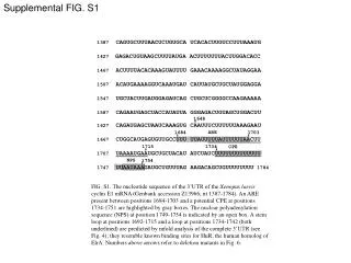
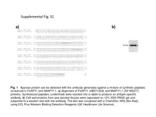
Supplemental FIG. S1 • 1387 CAGUGCUUUAACUCUGUGCA UCACACUUGUCCUUUAAAUG • 1427 GAGACUGUAAGCUUUUAUGA ACUUUUUUUACUUGGACACC • 1467 ACUUUUACACAAAGUAUUUU GAAACAAAAGGCUAUAGGAA • 1507 ACAUGAAAAGGUCAAAUGAU CAUUAUGCUGCUAUGGAGGA • 1547 UGCUACUUGAUGGAGAUCAG CUGCUCGGGGCCAAGAAAAA • 1587 CAGAAUGAGCUACCAUAUUA GGGAGACUGUAGCUGGACUU • 1648 • 1627 CAGAUGAGCUAAUCAAAGUG CAAUUUCUUUUUUAAAGAAU • 1684 1703 • 1667 CUGGCAUGAGUGUUGCCUUU UUAUUUUUAUUUUUUAACUU • 1715 1734 • 1707UAAAAUGAAUGCUGCUACAU AUCUAUCUUUUUUUUUUUUU • 1754 • 1747 UUAAUAAAGAUGCUGUUUAG AAGACAGCUGUUUUUUUU 1784 ARE CPE NPS FIG. S1. The nucleotide sequence of the 3’UTR of the Xenopus laevis cyclin E1 mRNA (Genbank accession Z13966; nt 1387-1784). An ARE present between positions 1684-1703 and a potential CPE at positions 1734-1751 are highlighted by gray boxes. The nuclear polyadenylation sequence (NPS) at position 1749-1754 is indicated by an open box. A stem loop at positions 1692-1715 and a loop at positions 1734-1742 (both underlined) are predicted by mfold analysis of the complete 3’UTR (see Fig. 4); they resemble known binding sites for HuR, the human homolog of ElrA. Numbers above arrows refer to deletion mutants in Fig. 6.


![[Fig. S1]](https://cdn3.slideserve.com/6448662/slide1-dt.jpg)