Acetylation Sites Identification in HDAC6: Mass Spectrometry Analysis and Conservation Alignments
This study identified acetylation sites in HDAC6 through mass spectrometry analysis of in vitro acetylated HDAC6, revealing conservation clusters among different species. Histone deacetylase activity of wild type and mutant HDAC6 proteins was evaluated.

Acetylation Sites Identification in HDAC6: Mass Spectrometry Analysis and Conservation Alignments
E N D
Presentation Transcript
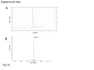
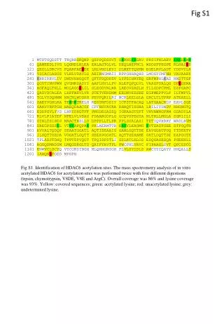
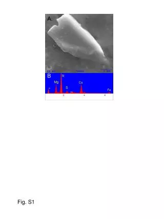
Fig S1 Fig S1. Identification of HDAC6 acetylation sites. The mass spectrometry analysis of in vitro acetylated HDAC6 for acetylation sites was performed twice with five different digestions (trpsin, chymotrypsin, V8DE, V8E and ArgC). Overall coverage was 86% and lysine coverage was 93%. Yellow: covered sequences; green: acetylated lysine; red: unacetylated lysine; grey: undetermined lysine.
B A Fig S2 * C D E Fig S2. HDAC6 alignementbetween species. Acetylation clusters A,B,C and D are more conserve between cattle, mouse and human HDAC6 than cluster E . The red star shows high conservation site, the blue star shows low conservation site.
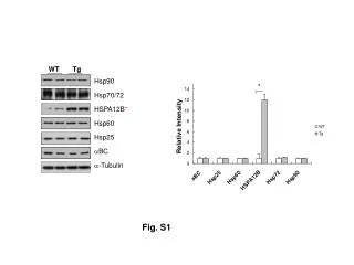
Fig S3 A B Deacetylase Activity (CMP) Fig S3. A. Recombinant HDAC6 and mutant proteins purified from baculovirus infected insect cells. B. Histone deacetylase activity of wild type and mutant HDAC6 proteins.


![[Fig. S1]](https://cdn3.slideserve.com/6448662/slide1-dt.jpg)