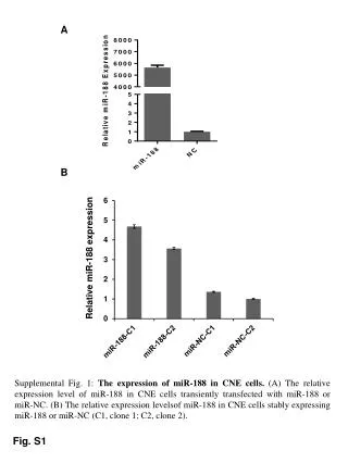
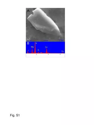
Fig. S1
3.74%. 3.54%. 45.60%. 41.22%. 3.54%. 3.74%. 47.21%. 43.34%. 4.35%. 5.15%. 2.01%. 3.00%. 4.35%. 5.15%. 33.84%. 34.73%. ROS dye added after trypsinization. ROS dye added before trypsinization. Percentage of ethidium fluorescence positive cells (O 2 .- production). Control. No GP.
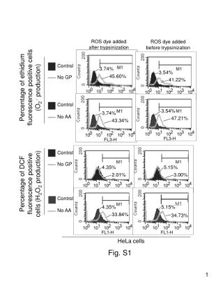

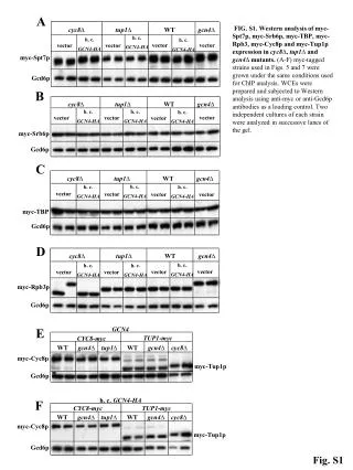
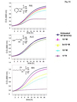
Fig. S1
E N D
Presentation Transcript
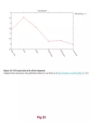
3.74% 3.54% 45.60% 41.22% 3.54% 3.74% 47.21% 43.34% 4.35% 5.15% 2.01% 3.00% 4.35% 5.15% 33.84% 34.73% ROS dye added after trypsinization ROS dye added before trypsinization Percentage of ethidium fluorescence positive cells (O2.- production) Control No GP Control No AA Percentage of DCF fluorescence positive cells (H2O2 production) Control No GP Control No AA HeLa cells Fig. S1
a Ethidium, control Ethidium, GP starvation DCF, control DCF, GP starvation U87 Cells b Ethidium, control Ethidium, AA starvation DCF, control DCF, AA starvation U87 Cells Fig. S2
LC3-I LC3-II β-Actin Fig. S3
* p < 0.002 80 No 3-MA Plus 3-MA 60 % Cells with AVOs or GFP-LC3 dots 40 20 0 a * * * * Fig. S4
Key Name M1 (%) Key Name M1 (%) Control 5.16 Control 5.16 No GP 45.13 36.52 No AA Wort 5.05 Wort+No AA 6.01 Wort+No GP 10.80 HeLa wt HeLa wt b (i) (ii) (iii) # p>0.05 * p<0.04 * # * * # # * * # HeLa wt HeLa SOD2 Fig. S4
Control siRNA * p<0.01 compared to “control siRNA” beclin-1 siRNA atg-7 siRNA c * * * * * * * * Fig. S4
SOD2 COX2 Actin Fig. S5
* p<0.05 * * * * * * Fig. S6
* * p<0.05 p<0.05 * * a * * b * * * * Fig. S7
50 40 30 % Cells with GFP-LC3 dots 20 10 0 LC3-I LC3-II β-actin p<0.01 * c * * * * d 10.4 28.5 45 38.5 71.2 63.8 14.2 0.007 2 0.8 4.1 0.1 Fig. S7
100 100 104 104 FL1-H FL3-H 1 mM H2O2 Treatment Control 200 200 Control Counts Counts 0 0 HEK293 cells LC3-I LC3-II β-Actin a Ethidium fluorescence DCF fluorescence b Fig. S8


![[Fig. S1]](https://cdn3.slideserve.com/6448662/slide1-dt.jpg)