Download
1 / 32
320 likes | 391 Views
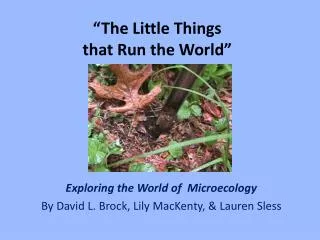
Explore the intriguing world of microecology with this detailed guide by David L. Brock, Lily MacKenty, and Lauren Sless. Learn how to extract, dilute, identify, and test soil samples to uncover the hidden life within. Discover the process of protozoa extraction and how to determine density vs. diversity of microorganisms. From chemical testing to acknowledging contributors, this book provides a comprehensive insight into the vital role of microbes in our ecosystem.

E N D
“The Little Thingsthat Run the World” Exploring the World of Microecology By David L. Brock, Lily MacKenty, & Lauren Sless
Sample Collecting • use soil cylinders 10-15 cm deep; keep in fresh plastic bags (don’t reuse to avoid contamination) • should collect min. of 3 samples from each location • make sure to collect all samples on the same day & time • remember: soil is still “alive” in the plastic bag
Protozoa Extraction • be sure to use distilled water and not tap water; but it does not need to be sterile • filter the Uhlig run-off a second time for improved viewing • methyl green is the preferred stain • to quantify: [(# per field of view at 40X) • (total ml of water used) • 747] (grams of sifted soil ) = # of protozoa per gram of soil
Step 1: collect and label clean petri dishes for each soil sample
Step 2b: then put dried soil into a small cup and cover with a 1 mm2 nylon mesh; sift 10 g into a clean petri dish
Step 4: allow sample(s) to sit for 7 hours at room temperature
Step 5: make a modified Uhlig ciliate sandy sediment separator(s) out of plastic cups & 2 sheets of nylon bridal veil
Step 6: add 30 ml of distilled water to the bottom of a 100x15 mm petri dish
Step 7: place Uhlig extractor into petri dish & scoop the rehydrated soil sample into the extractor. Allow to filter for 24 hrs at room temp.
Step 8: filter the sample a second time using qualitative filter paper
Step 9a: prepare microscope slides for viewing from the second filtrate
Step 9b: using a capillary tube, add methyl green dye to a microscope slide (1 ul = 1 drop from the tube)
Step 9c: add 18 ul of the filtrate using a graduated Beral-type pipette (the first demarcation) and cover with an 18 x 18 mm2 cover slip
Step 10a: To prevent slides from drying out, dip the tip of a flat toothpick into petroleum jelly. Step 10b: Then carefully streak the jelly along the edges of the cover slip to create a seal. Slide can now be observed at leisure up to 3 hours.
Nine Fields of View Types of Protozoa Ciliophora Shelled Amoeba Mastigiophora Sporozoa Non-shelled Amoeba • Protozoa will appear translucent and slightly bluer than their surroundings. Make sure to adjust the light so that the maximum number of protozoa that are visible, and if needed use a counter.
Serial Dilutions • use for bacteria, yeast, and mold counts in cfu/cm3; formula = # of colonies • 102 • 10 |dilution factor| • be sure to use sterile water (boiled & cooled works perfectly fine) • can reuse dilution tubes but clean thoroughly • easily adapted to “low-tech” with disposable graduated plastic droppers
Step1: place a 1 cc sample of soil along with 10 ml of sterile water into a 15 ml transformation tube; cap, shake vigrously, and remove 1 ml of the soil/water mixture to add to a second transformation tube containing 9 ml of water.
Step 2: repeat, placing 1 ml from 2nd tube into a 3rd tube, and so on until sample is diluted at least 4 times
Step 4: plate the 100 µl samples with media & method of your choice
Step 6: examine each plate from a dilution series to find the ones with between 5 & 30 colonies; count the colonies on only those plates & use the formula to calculate the density of bacteria in the original cc of each soil sample
Chemical Testing • goggles & gloves! • always test at same time you sample microbe populations • pH, calcium, nitrate, & phosphate show nice relationships with protozoa as well as bacteria • active iron, aluminum, & manganese provide a good challenge for your better students
Acknowlegements • Kate Brockmeyer, Katie Loya, & Mariel Torres • Institute for Ecosystem Studies • ReliaStar/Northern Life “Unsung Heroes” Program • Toshiba America Foundation • Human Capital Development, Inc • Paul F-Brandwein Institute • National Science Foundation • Gustav Ohaus Awards • Captain Planet Foundation, Inc. • Waksman Foundation for Microbiology • The Josowitz Family • Flinn Scientific • SeaWorld/Busch Gardens/Fujifilm Environmental Excellence Awards
For further information: --visit the program web page athttps://faculty.rpcs.org/brockda & click on the “Little Things” link,--visit the Environmental Science Summer Research Experience for Young Women athttps://faculty.rpcs.org/essre,--or e-mail brockda@rpcs.org