EEG / MEG: Experimental Design & Preprocessing
EEG / MEG: Experimental Design & Preprocessing. Ioannis Sarigiannidis Wen-Jing Lin. Outline. Experimental Design fMRI M/EEG A nalysis Oscillatory activity EP Design Inferences Limitations Combined Measures. Preprocessing in SPM8 Data Conversion Montage Mapping Epoching


EEG / MEG: Experimental Design & Preprocessing
E N D
Presentation Transcript
EEG / MEG: Experimental Design & Preprocessing Ioannis Sarigiannidis Wen-Jing Lin
Outline Experimental Design • fMRI M/EEG • Analysis • Oscillatory activity • EP • Design • Inferences • Limitations • Combined Measures Preprocessing in SPM8 • Data Conversion • Montage Mapping • Epoching • Downsampling • Filtering • Artefact Removal • Referencing
MEG vs. EEG Signal from pyramidal neurons of the cortex
MEG is mostly sensitive to tangential fields gyrus sulcus
Two types of MEG/EEG analysis Event related changes (EP / ERP – ERF) Oscillatory activity – cortical rhythms (Time-frequency analysis) Otten, L. (2012, November 21). EEG/MEG Acquisition, Analysis and Interpretation, MSc Cognitive Neuroscience, UCL
Oscillations Otten, L. (2012, November 21). EEG/MEG Acquisition, Analysis and Interpretation, MSc Cognitive Neuroscience, UCL
Evoked vs. Induced (Hermann et al. 2004)
Oscillations • Delta (0 – 4 Hz) • Large-scale cortical integration • Attentional and syntactic language processes • Deep sleep • Theta (4 – 8 Hz) • Codes locations in space, navigation • Declarative memory processes • Successful memory encoding • Episodic memory processing
Oscillations • Alpha (8 – 12or 13 Hz) • Closed eyes • Level of cortical activation • Cortical and behavioral deactivation or inhibition • Perceptual, memory and attentional processes • Beta (12 – 30 Hz) • Alert, REM sleep • Attention, and higher cognitive function • Stop movement
Oscillations • Gamma (30 – 80 Hz) • Visual awareness • Binding of information • Encoding, retention and retrieval of information independent of sensory modality • Recording gamma activity in the human EEG is difficult • very small amplitude • similarity in terms of its frequency characteristics with electrical muscle activity • microsaccades – confused with gamma
Oscillations • Non-averaged data collected during continuous stimulation or task performance (or during rest) lends itself to analysis of spectral power. • i.e. We can do Fourier analysis and look at spectra (not-event related – break data in arbitrary segments and do some averaging) • e.g. sleep studies, mental states (e.g. meditation)
EP vs. ERP / ERF • Evoked potential (EP) • short latencies (< 100ms) • small amplitudes (< 1μV) • sensory processes • Event related potential / field • longer latencies (100 – 600ms), • higher amplitudes (10 – 100μV) • higher cognitive processes but used interchangeably in general
ERP/ ERF Average potential/ field at the scalp relative to some specific event Stimulus/ Event Onset Baseline: typically 100ms before the onset of the stimulus
ERP/ ERF Non-time locked activity(noise) lost via averaging Averaging
Experimental design • Number of trials • EP: 120 trials, 15-20% will be excluded • Oscillatory activity: 40-50 trials • Duration of stimuli / task • Short: Averaged EP is fine • (Very) long: spectrotemporal analysis on averaged EP or non-averaged data • Collecting Behavioral Responses • Only if necessary!
Inferences Not Based On Prior Knowledge Observe • Time course • Amplitude • Distribution across scalp • Differences in ERP Infer • Timing • Degree of engagement • Functional equivalence of underlying cognitive process
Inferences Based On Prior Knowledge An “ERP component is scalp-recorded electrical activity that is generated in a given neuroanatomical module when a specific computational operation is performed.” (Luck 2004, p. 22)
Observed vs. Latent Components Observed waveform Latent components OR
Design Strategies • Focus on specific, large and easily isolated component • E.g., P3, N400, LRP, N2pc… • Use well-studied experimental manipulations • Isolate components with different waves • Component-independent experimental designs
Design Strategies • Avoid confounds and misinterpretations • Physical stimulus confounds • Side effect • What you manipulated indirectly influences other things • Vary conditions within rather than between blocks • Be cautious of behavioral confounds
Sources of Noise in EEG • EEG activity not elicited by stimuli • e.g. alpha waves • Trial-by-trial variations • Articfactual bioelectric activity • eye blinks, eye movement, muscle activity, skin potentials • Environmental electrical activity • e.g. from monitors
Signal-to-Noise Ratio • Size of the noise in average = (1/√N) ×R • Number of trials: • Large component: 30– 60 per condition • Medium component: 150– 200 per condition • Small component: 400– 800 per condition • Double with children or psychiatric patients
Limitations • Ambiguous relation between observed ERP and latent components • Signal distorted en route to scalp • arguably worse in EEG than MEG (head as “spherical conductor”) • MEG: application restrictions • patients with implants • Poor localization (cf. “inverse problem”)
Combining Techniques-How? • Converging evidence • Combination of different information from different experiments • Generative models • Establish generative models for which parameters are estimated from data of different nature.
Combining Techniques - Why? • fMRI & M/EEG • BOLD activity can occur without M/EEG. • Specific spatial configurations of the cells or of the sources may annihilate signals at the surface of the scalp. • M/EEG activity can occur in the absence of BOLD • synchronization may not necessarily consume enough energy to be seen in BOLD. • The two activities are not necessarily spatially congruent. Many studies have found discrepancies between EEG dipolar localization and fMRI • M/EEG increased resolution improved localization
Technical M/EEG Considerations • Amplifier and filter settings • Sampling frequency • EEG • Number, type, location of electrodes • Reference electrodes • MEG • equipment and participant compatible with MEG? • [digitize 3D head] matched to [structural MRI]
Outline Experimental Design • fMRI M/EEG • Analysis • Oscillatory activity • EP • Design • Inferences • Limitations • Combined Measures Preprocessing in SPM8 • Data Conversion • Montage Mapping • Epoching • Downsampling • Filtering • Artefact Removal • Referencing
PREPROCESSING • Raw data to averaged ERP (EEG) or ERF (MEG) using SPM 8
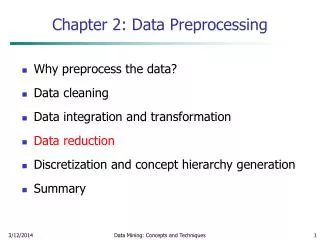
Conversion of data • Convert data from its native machine-dependent format to MATLAB based SPM format • Do not define setting: • “Just read” • Define settings: • Read data as continuous or as trials • Select channels • Define file name *.mat (data) *.bdf *.bin *.eeg *.dat (other info)
Downsampling • Sampling frequency: number of samples per second taken from a continuous signal • Data are usually acquired with a very high sampling rate • SF should be greater than twice the maximum frequency of the signal of interest? • Downsamplingreduces the file size and speeds up the subsequent processing steps (e.g. 200 Hz)
Montage and Referencing • Identify vEOG and hEOG channels, remove several channels that don’t carry EEG data • Specify reference for remaining channels • Single electrode reference: free from neural activity of interest • Average reference: Output of all amplifiers are summed and averaged and the averaged signal is used as a common reference for each channel
Epoching • Cut out chunks of continuous data (= single trials) • Specify time window associated with triggers [prestimulus time, poststimulus time] • Baseline-correction: automatic; the mean of the prestimulus time is subtracted from the whole trial • Segment length: at least 100 ms for baseline-correction; the longer the more artefacts • Padding: adds time points before and after each trial to avoid ‘edge effects’ when filtering For multisubject/batch epoching in future
Filtering • EEG data consist of signal and noise • Some noise is sufficiently different in frequency content from the signal. It can be suppressed by attenuating different frequencies. • Non-neural physiological activity (skin/sweat potentials); (drifts – high pass filter takes care of that) noise from electrical outlets (bandstop) • SPM8: Butterworth filter • High-, low-, band- pass or bandstop filter • Any filter distorts at least some part of the signal
Artefact Removal • Eye movements • Eye blinks • Muscle activity • Skin potentials • ‘Boredom’ (alpha waves) • Headmovements
ArtefactRemoval • Removal • Hand-picked • Automatic SPM functions: • Thresholding (e.g. 200 μV) • 1st– bad channels, 2nd – bad trials • No change to data, just tagged • Robust averaging: estimates weights (0-1) indicating how artefactual a trial is
References • Ashburner, J. et al. (2010). SPM8 Manual. http://www.fil.ion.ucl.ac.uk/spm/ • Hansen, C.P., Kringelbach M.L., Salmelin, R. (2010) MEG: An Introduction to Methods. Oxford University Press, • Hermann, C. et al. (2004). Cognitive functions of gammaband activity: memory match and utilization. Trends in Cognitive Science, 8(8), 347-355. • Herrmann, C. S., Grigutsch, M., & Busch, N. A. (2005). EEG oscillations and wavelet analysis. In T. C. Handy (Ed.), Event-related potentials: A methods handbook (pp. 229-259). Cambridge, MA: MIT Press. • Luck, S. J. (2005). Ten simple rules for designing ERP experiments. In T. C. Handy (Ed.), Event-related potentials: a methods handbook. Cambridge, MA: MIT Press. • Luck, S. J. (2010). Powerpoint Slides from ERP Boot Camp Lectures. http://erpinfo.org/Members/ldtien/bootcamp-lecture-pptx • Otten, L. (2012, November 21). EEG/MEG Acquisition, Analysis and Interpretation, MSc Cognitive Neuroscience, UCL • Otten, L. J. & Rugg, M. D. (2005). Interpreting event-related brain potentials. In T. C. Handy (Ed.), Event-related potentials: a methods handbook. Cambridge, MA: MIT Press.. • Sauseng, P., & Klimesch, W. (2008). What does phase information of oscillatory brain activity tell us about cognitive processes? [Review]. Neuroscience and Biobehavioral Reviews, 32(5), 1001-1013. doi: 10.1016/j.neubiorev.2008.03.014 • MfD slides from previous years
Kilner, unpublished Wager et al. Neuroimage, 2005