Introduction to the Microscope
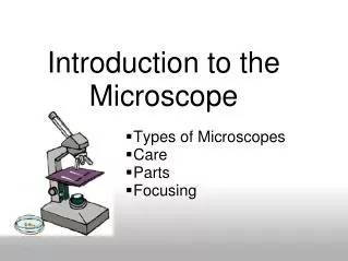
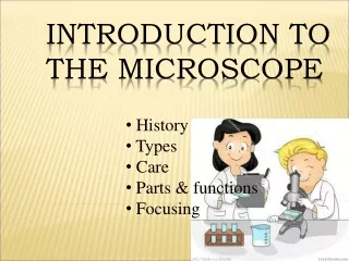
Introduction to the Microscope. History Types Care Parts Focusing. Who invented the microscope?. Diggs? Hans and Zcharias Janssen?. Who really used the microscope?. Antony van Leeuwenhook. Robert Hooke. He figured out that living things are made of cells. Types of Microscopes.

Introduction to the Microscope
E N D
Presentation Transcript
Introduction to the Microscope History Types Care Parts Focusing
Who invented the microscope? Diggs? Hans and Zcharias Janssen?
Who really used the microscope? Antony van Leeuwenhook
Robert Hooke He figured out that living things are made of cells.
Types of Microscopes Compound -More than one lens -Has own light -Light must see through the sample
Types of Microscopes Stereomicroscope/Dissecting -Do not need light to pass through object -Low magnification
Types of Microscopes Electron -Very powerful -Passes electrons through an object rather than light -Coat with gold or platinum to attract particles
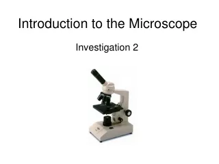
Label the parts on your microscope picture. Eyepiece/ocularlensmagnification = 10X
Label the parts on your microscope picture. Eyepiece/ocularlensmagnification = 10X Arm-Support
Label the parts on your microscope picture. Eyepiece/ocularlensmagnification = 10X Arm-Support Stage - support
Label the parts on your microscope picture. Eyepiece/ocularlensmagnification = 10X Arm-support Stage - support
Label the parts on your microscope picture. Eyepiece/ocularlensmagnification = 10X Arm-support Stage - support Coarse adjust – general focus Fine Focus –medium and high power focus
Label the parts on your microscope picture. Eyepiece/ocularlensmagnification = 10X Arm-Support Stage - support Coarse adjust – general focus Fine Focus – medium andhigh power focus Base -support
Label the parts on your microscope picture. Eyepiece/ocularlensmagnification = 10X Arm-Support Stage - support Coarse adjust – general focus Fine Focus –high power focus Light source Base -support
Label the parts on your microscope picture. Eyepiece/ocularlensmagnification = 10X Arm-Support Stage - support Coarse adjust – general focus Diaphragm – adjusts amount of light Fine Focus –high power focus Light source Base -support
Label the parts on your microscope picture. Eyepiece/ocularlensmagnification = 10X Arm-Support Stage - support Stage clips – holds slide Coarse adjust – general focus Diaphragm – adjusts amount of light Fine Focus –high power focus Light source Base -support
Label the parts on your microscope picture. Eyepiece/ocularlensmagnification = 10X Arm-Support Objective Lenses Stage - support Stage clips – holds slide Coarse adjust – general focus Diaphragm – adjusts amount of light Fine Focus –medium and high power focus Light source Base -support
Label the parts on your microscope picture. Body Tube Eyepiece/ocularlensmagnification = 10X Revolving nosepiece Arm-Support Objective Lenses Stage - support Stage clips – holds slide Coarse adjust – general focus Diaphragm – adjusts amount of light Fine Focus –high power focus Light source Base -support
Eyepiece Objective Lens Magnification Low Power 10X 4X 40X Medium Power 10X 10X 100X High Power 10X 40X 400X • Magnification of a Compound Microscope • Because you are looking through multiple lenses the lenses have a “compounding” effect. • The eyepiece always magnifies 10X
FIELD OF VIEW The circular area viewed through the eyepiece.Magnification – The number of times the image of an object is increased insize by a lens system
MAGNIFICATION The number of times the image of an object is increased in size by a lens system
Microscope Care Always carry with 2 hands – one on the base and the other on the arm Do not force knobs Click Nosepiece to the lowest (shortest) setting when done – otherwise known as the park position Cover the microscope when done
Using the Microscope • Place the Slide on the Microscope • Use Stage Clips • Click Nosepiece to the lowest (shortest) setting • Look into the Eyepiece • Use the Coarse Focus
Using Medium and High Power Follow steps to focus using low power Click the nosepiece to the medium or longest objective Bring the stage as close as possible to the objective lens using the Coarse Focusing Knob Use the Fine Focus Knob to bring the slide into focus