Download
1 / 37
470 likes | 1.05k Views
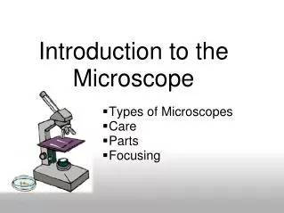
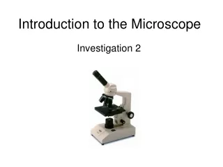
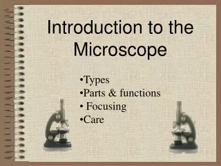
Introduction to the Microscope. Types Parts & functions Focusing Care. Types of Microscopes. Dissection Microscope Scanning Electron Microscope (SEM) Transmission Electron Microscope (TEM) Compound Light Microscopes. Dissection Microscope.

E N D
Introduction to the Microscope • Types • Parts & functions • Focusing • Care
Types of Microscopes • Dissection Microscope • Scanning Electron Microscope (SEM) • Transmission Electron Microscope (TEM) • Compound Light Microscopes
Dissection Microscope A dissection microscope is light illuminated. The image that appears is three dimensional. It is used for dissection to get a better look at the larger specimen. You cannot see individual cells because it has a low magnification. (also called stereo microscope)
Dissection Microscope Images Head of a moth pupa 60x Sunflower with moth pupa in the stem 10x
Scanning Electron Microscope - SEM SEM use electron illumination. The image is seen in 3-D. It has high magnification and high resolution. The specimen is coated in gold and the electrons bounce off to give you and exterior view of the specimen. The pictures are in black and white.
Scanning Electron Microscope Images pigeon blood cockroach antenna
Transmission Electron Microscope - TEM TEM is electron illuminated. This gives a 2-D view. Thin slices of specimen are obtained. The electron beams pass through this. It has high magnification and high resolution.
Transmission Electron Microscope Images bacillus bacteria dividing mitochondrion
Compound Microscope Compound microscopes are light illuminated. The image seen with this type of microscope is two dimensional. This microscope is the most commonly used. You can view individual cells, even living ones. It has high magnification. However, it has a low resolution.
Compound Microscope Images Paulownia Wood c.s.200x Frog’s blood 1,000x
Compound Light Microscope Light Microscope - the models found in most schools, use compound lenses to magnify objects. The lenses bend or refract light to make the object beneath them appear closer. Common magnifications: 40x, 100x, 400x
How a Microscope Works Convex Lenses are curved glass used to make microscopes (and glasses etc.) Convex Lenses bend light and focus it in one spot.
How a Microscope Works Ocular Lens (Magnifies Image) Objective Lens (Gathers Light, Magnifies And Focuses Image Inside Body Tube) Body Tube (Image Focuses) • Bending Light: The objective (bottom) convex lens magnifies and focuses (bends) the image inside the body tube and the ocular convex (top) lens of a microscope magnifies it (again).
Microscope Parts Ocular lens BodyTube RevolvingNosepiece Arm ObjectiveLens Stage StageClips Coarse adjustment knob Diaphragm Fineadjustment knob Light Base
ocular lens Ocular lens magnifies; where you look through to see the image of your specimen. They are usually 10X or 15X power.
arm supports the tube and connects it to the base arm
stage the flat platform where you place your slides stage
coarse adjustment knob moves stage (or body tube) up and down coarse adjustment knob
fine adjustment knob small, round knob on the side of the microscope used to fine-tune the focus of your specimen after using the coarse adjustment knob fine adjustment knob
base the bottom of the microscope, used for support base
body tube connects the eyepiece to the objective lenses body tube
revolving nosepiece the part that holds two or more objective lenses and can be rotated to easily change power revolving nosepiece
objective lenses Adds to the magnification Usually you will find 3 or 4 objective lenses on a microscope. They almost always consist of 4X, 10X, 40X and 100X powers. objective lens
stage clips Stage clips hold the slides in place. stage clips
diaphragm controls the amount of light going through the specimen diaphragm
light makes the specimen easier to see light
Eyepiece BodyTube RevolvingNosepiece Arm ObjectiveLens Stage StageClips CoarseFocus Diaphragm FineFocus Light Base
The Light Microscope Guidelines for Use Always carry with 2 hands Only use lens paper for cleaning Do not force knobs Always store covered Always go from small to large magnification Course adjustment first Except on 100x mag.
Focusing Specimens 1. Always start with the scanning objective. Odds are, you will be able to see something on this setting. Use the Coarse Knob to focus and then the fine adjustment knob until clear, image may be small at this magnification, but you won't be able to find it on the higher powers without this first step. Do not use stage clips, try moving the slide around until you find something.
2. Once you've focused on Scanning, switch to Low Power. Use the Coarse Adjustment Knob to refocus. Then use the Fine Adjustment Knob to make the image crystal clear. Again, if you haven't focused on this level, you will not be able to move to the next level. 3. Now switch to High Power. (If you have a thick slide, or a slide without a cover, do NOT use the high power objective). At this point, ONLY use the Fine Adjustment Knob to focus specimens. Recap 1. Scanning --> use coarse and fine knob 2. Low power --> use coarse and fine knob 3. High power --> use fine knob only DO NOT SKIP STEPS!!!!
Your slide MUST be focused on low power before attempting this step Click the nosepiece to the longest objective Do NOTuse the Coarse Focusing Knob, this could crack the slide or the lens Use the Fine Focus Knob to bring the slide
Drawing Specimens 1. Use pencil - you can erase and shade areas 2. All drawings should include clear and proper labels (and be large enough to view details). Drawings should be labeled with the specimen name and magnification. 3. Labels should be written on the outside of the circle. The circle indicates the viewing field as seen through the eyepiece, specimens should be drawn to scale - ie..if your specimen takes up the whole viewing field, make sure your drawing reflects that.
MagnificationMost microscopes have 3 magnifications: Scanning, Low and High. Each objective will have written the magnification. In addition to this, the ocular lens (eyepiece) has a magnification. The total magnification is the ocular x objective
Storing your Microscope • Stage all the way down • Light turned off • Cord wrapped correctly • Covered • The smallest objective lens should be in place • Carry it by the neck and the base
Quiz Over the Microscope 1. When focusing a specimen, you should always start with the ___________________ objective. 2. When using the high power objective, only the ___________ knob should be used. 3. The type of microscope used in most science classes is the _________________ microscope 4. What part of the microscope can adjust the amount of light that hits the slide? ______________________________
5. You should carry the microscope by the ________ and the __________. 6. The objectives are attached to what part of the microscope (it can be rotated to click the lenses into place): _______________ ________________ 7. You should always store you microscope with the ________________ objective in place. 8. A microscope has an ocular objective of 10x and a high power objective of 50x. What is this microscope's total magnification? ____________