Download
1 / 43
990 likes | 4.41k Views
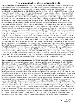
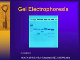
Two dimensional gel electrophoresis. What is 2D gel electrophoresis?. Separation and identification of proteins in a sample by displacement in 2 dimensions oriented at right-angle to one another. First dimension: Isoelectric focussing Second dimension: SDS PAGE.

E N D
What is 2D gel electrophoresis? • Separation and identification of proteins in a sample by displacement in 2 dimensions oriented at right-angle to one another. • First dimension: Isoelectric focussing • Second dimension: SDS PAGE
Basis of 2D gel electrophoresis • Highly unlikely for proteins to be similar in two distinct properties • Isoelectric point • Protein mass in SDS PAGE • Protein complex mass in native PAGE
Advantages of 2D gel electrophoresis • Resolution : 104 - 105 proteins from a sample. • Preset conditions ( pH ranges, size of the gel, staining methods etc) can be changed to increase resolution. • Delivers a map of intact protein that can be stored and analyzed at will. One of the core technology of proteomics.
Limitations • Hard to resolve • very acidic or very basic proteins • very low or very high molecular weight. • Poor resolution • hydrophobic or membrane bound • nuclear proteins • Sample size • proteins cannot be amplified like DNA. • Difficulty in process automation
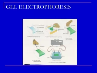
2D gel electrophoresis steps • Sample preparation • First dimension separation • Isoelectric focussing • Second dimension separation • SDS PAGE • Detection of spots • Analysis of protein spots
Sample preparation • Must remove substances that might interfere with separation process such as salts, polar detergents (SDS), lipids, polysaccharides, nucleic acids • Must try to keep proteins soluble during both phases of electrophoresis process
Sample preparation A. Contaminants and their removal procedure
Sample preparation B. Solubilise proteins: Lysis Solution Composition CHAPS: 3-[(3-Cholamidopropyl)dimethylammonio]-1-propanesulfonate DTT: Dithrioerythritol; DTE: epimer of DTT
Isoelectric focussing • Separation on basis of pI, not Mw • Requires very high voltages (5000V) • Requires a long period of time (10h) • Presence of a pH gradient is critical • Uses ampholytes to establish pH gradient
NH3+ NH3+ NH2 COOH COO- COO- NH3+ NH3+ NH2 COOH COO- COO- pH < pI Net positive charge pH = pI pH > pI Net negative charge Isoelectric focussing Isoelectric point is the pH of a solution at which the net charge of protein is zero. In electrophoresis there is no motion of the particles in an electric field at the isoelectric point.
Isoelectric focussing pH gradients • Soluble Ampholytes • heterogeneous polymers of charged compounds in a regular (tube) polyacrylamide gel • Variability: batch to batch • pH range about 4-8 at equilibrium • Immobilised pH gradient • Acrylamide derivatives with charged side chains immobilised on a strip • Stable and reproducible gradient • Higher mechanical strength • Higher protein loading capacity
10 3 Broad range 7 4 Medium range 11 6 3.5 4.5 5.5 6.7 Narrow range 4.0 5.0 6.0 Isoelectric focussing IPG strips (3 mm x 18 cm x 0.5 mm)
Isoelectric focussing • Load sample per groove • Peel off protective film from strip • Place IPG strip gel facing down in groove • Rehydrate overnight (~22 hrs) at room temperature
Isoelectric focussing IEF run Slowly increase the voltage and apply a high voltage at end to obtain sharp narrow zones of protein
Isoelectric focussing • Equilibration • Reduction : • 50mM DTT in equilibration buffer for 15 minutes • Maintains proteins in a reduced state • Alkylation (optional): • 125mM iodoacetamide in equilibration buffer for 15 minutes • Alkylating sulfhydryl group reduces vertical streaking Equilibration buffer: 6M urea, 30% glycerol; 1.6% SDS; 0.002% bromophenol blue; 45 mM Tris base (pH 7.0)
SDS-PAGE • Second dimension SDS-PAGE steps • Equilibrating IPG strips after IEF. • Applying IPG strips to the second dimension SDS gel. • Performing SDS-PAGE
- + SDS-PAGE: Step1 After IEF run • Remove the IPG strip from the tray • Place IPG strip facing up in the equilibration buffer
SDS-PAGE SDS-PAGE Marker in paper 0.5% agarose in running buffer - + SDS-PAGE SDS-PAGE: Step 2 IPG strip in Equilibration buffer IPG strip is placed on top of pre-cast SDS-PAGE gel and electric current applied
SDS-PAGE: Step 3 • Separation on basis of MW, not pI • Requires modest voltages (200V) • Requires a shorter period of time (2h) • Presence of SDS is critical to disrupting structure and making mobility • Degree of resolution determined by %acrylamide & electric field strength
Detection Coommassie stain Silver stain
Detection Copper stain SYPRO flourescent
Vertical streaks Insufficient equilibration, insufficient SDS Horizontal streaks Sample not completely solubilized prior to application on IPG, sample poorly soluble in rehydration solution, ionic impurities, ionic detergents Trouble shooting
Analysis of spots • 2D gel software
Analysis of spots • Commercial Software • Melanie 4 (GeneBio - Windows only) http://ca.expasy.org/melanie • ImageMaster 2D Elite (Amersham) http://www.imsupport.com/ • Phoretix 2D Advanced http://www.phoretix.com/ • PDQuest 6.1 (BioRad - Windows only) http://www.proteomeworks.bio-rad.com/html/pdquest.html
Analysis of spots • Common Software Features • Image contrast and coloring • Gel annotation (spot selection & marking) • Automated peak picking • Spot area determination (Integration) • Matching 2 gels • Stacking/Aligning/Comparing gels
Analysis • Excision the spots of interest • Pick up the protein gel spot from gel • Manual • Automatic • In-gel digestion: • Washing process • Dehydratation and drying • Trypsin digestion (50 ng trypsin, 37C 16h) • Extraction • Desalt and concentrate the peptide
Analysis • Identification of eluted protein spots • MALDI-TOF (Matrix Assisted Laser Desorption/Ionization-Time Of Flight) • MS (Mass Spectrometry), • Peptide mass fingerprint
Applications • Analyzing proteome profiles • Detecting post- or co-translational modifications • Discovering new drug targets • Studying protein expression in normal, disease, or developmental states • Identifying novel proteins
Clinical specimens Cryostat Laser-captured microdissector (LCM) Normal cells Tumor cells 2D gel electrophoresis Immage system SDS-PAGE isoelectrofocusing (?????)
Schedule of a 2D Experiment Day 1: Sample preparation and IEF 1. Load protein sample onto IPG strip (IEF) 2. Run the IEF (about 24 hours) 3. Polyacrylamide gel casting Day 2: Equilibrium IPG strip and running SDS-PAGE 1. Remove IPG strip from IEF machine 2. Equilibrium IPG strip 3. Put IPG strip onto SDS-PAGE 4. Run the SDS-PAGE (overnight)
Schedule of a 2D Experiment Day 3: Staining, image scanning and image analysis 1. Remove the gel from the cassette 2. Stain the gel by SYPRO Ruby or silver 3. Scan the gel image 4. Image analysis Day 4: In-gel digestion, MALDI-TOF and database search 1. Pick the protein gel spot from gel 2. In-gel digestion 3. Spot the sample onto MALDI chip 4. MALDI-TOF analysis 5. Database search
Online protocols • http://ca.expasy.org/ch2d/protocols/ • http://www.abdn.ac.uk/~mmb023/protocol.htm • http://www.aber.ac.uk/~mpgwww/Proteome/Tut_2D.html • http://www.noble.org/PlantBio/MS/protocols.html