Joints
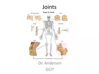
Joints. Chapter 8. Joints (Articulations). Articulation —site where two or more bones meet Functions of joints: Give skeleton mobility Hold skeleton together. Functional Classification of Joints . Based on amount of movement allowed by the joint Three functional classifications:

Joints
E N D
Presentation Transcript
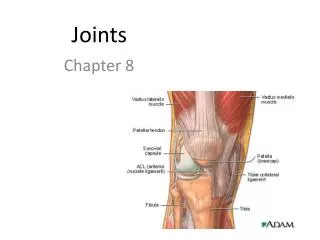
Joints Chapter 8
Joints (Articulations) • Articulation—site where two or more bones meet • Functions of joints: • Give skeleton mobility • Hold skeleton together
Functional Classification of Joints • Based on amount of movement allowed by the joint • Three functional classifications: • Synarthroses—immovable • Amphiarthroses—slightly movable • Diarthroses—freely movable
Structural Classification of Joints • Based on binding material and joint cavity • Three structural classifications: • Fibrous • Cartilaginous • Synovial
Fibrous Joints • Bones joined by dense fibrous connective tissue • No joint cavity • Most are synarthrotic (immovable) • Three types: • Sutures • Syndesmoses • Gomphoses
Fibrous Joints: Sutures • Rigid, interlocking joints containing short connective tissue fibers • Allow for growth during youth • In middle age, sutures ossify and are called synostoses
Fibrous Joints: Syndesmoses • Bones connected by ligaments • Movement varies from immovable to slightly movable • Examples: • Synarthrotic distal tibiofibular joint • Diarthroticinterosseous connection between radius and ulna
Fibrous Joints: Gomphoses • Peg-in-socket joints of teeth in alveolar sockets • Fibrous connection is the periodontal ligament
Cartilaginous Joints • Bones united by cartilage • No joint cavity • Two types: • Synchondroses • Symphyses
Cartilaginous Joints: Synchondroses • A bar or plate of hyaline cartilage unites the bones • All are synarthrotic
Cartilaginous Joints: Symphyses • Hyaline cartilage covers the articulating surfaces and is fused to an intervening pad of fibrocartilage • Strong, flexible amphiarthroses
Synovial Joints • All are diarthrotic • Include all limb joints; most joints of the body • Distinguishing features: • Articular cartilage: hyaline cartilage • Joint (synovial) cavity: small potential space • Articular (joint) capsule: outer fibrous capsule of dense irregular connective tissue, inner synovial membrane of loose connective tissue • Synovial fluid: viscous slippery filtrate of plasma + hyaluronic acid – lubricates and nourished articular cartilage
Ligament Joint cavity (contains synovial fluid) Articular (hyaline) cartilage Fibrous capsule Articular capsule Synovial membrane Periosteum Figure 8.3
Synovial Joints Distinguishing features cont: 5. Three possible types of reinforcing ligaments: • Capsular (intrinsic)—part of the fibrous capsule • Extracapsular—outside the capsule • Intracapsular—deep to capsule; covered by synovial membrane 6. Rich nerve and blood vessel supply: • Nerve fibers detect pain, monitor joint position and stretch • Capillary beds produce filtrate for synovial fluid
Synovial Joints: Friction-Reducing Structures • Bursae: • Flattened, fibrous sacs lined with synovial membranes • Contain synovial fluid
Synovial Joints: Friction-Reducing Structures • Tendon sheath: • Elongated bursa that wraps completely around a tendon
Stabilizing Factors at Synovial Joints • Shapes of articular surfaces (minor role) • Ligament number and location (limited role) • Muscle tone • Extremely important in reinforcing shoulder and knee joints and arches of the foot
Synovial Joints: Movement • Muscle attachments across a joint: • Origin—attachment to the immovable bone • Insertion—attachment to the movable bone • Muscle contraction causes the insertion to move toward the origin • Movements occur along transverse, frontal, or sagittal planes
Synovial Joints: Range of Motion • Nonaxial—slipping movements only • Uniaxial—movement in one plane • Biaxial—movement in two planes • Multiaxial—movement in or around all three planes
Synovial Joint Movement: • Gliding: one flat bone surface glides or slips over another similar surface • Examples: • Intercarpal joints • Intertarsal joints • Between articular processes of vertebrae
Synovial Joint Movement: • Angular: (1)movements that occur along the sagittal plane: • Flexion—decreases the angle of the joint • Extension— increases the angle of the joint • Hyperextension—excessive extension beyond normal range of motion
Synovial Joint Movement • Angular: (2)movements that occur along the frontal plane: • Abduction—movement away from the midline • Adduction—movement toward the midline • Circumduction
Synovial Joint Movement • Rotation: The turning of a bone around its own long axis • Examples: • Between C1 and C2 vertebrae • Rotation of humerus and femur
Synovial Joints: Special Movements • Movements of radius around ulna: • Supination (turning hand backward) • Pronation (turning hand forward) • Movements of the foot: • Dorsiflexion (upward movement) • Plantar flexion (downward movement)
Synovial Joints: Special Movements • Movements of the foot: • Inversion (turn sole medially) • Eversion (turn sole laterally) • Movements in a transverse plane: • Protraction (anterior movement) • Retraction (posterior movement)
Synovial Joints: Special Movements • Elevation (lifting a body part superiorly) • Depression (moving a body part inferiorly) • Opposition of the thumb • Movement in the saddle joint so that the thumb touches the tips of the other fingers
Classification of Synovial Joints • Six types, based on shape of articular surfaces: • Plane • Hinge • Pivot • Condyloid • Saddle • Ball and socket
Plane Joints • Nonaxial joints • Flat articular surfaces • Short gliding movements
Hinge Joints • Uniaxial joints • Motion along a single plane • Flexion and extension only
Pivot Joints • Rounded end of one bone conforms to a “sleeve,” or ring of another bone • Uniaxial movement only
Condyloid (Ellipsoidal) Joints • Biaxial joints • Both articular surfaces are oval • Permit all angular movements
Saddle Joints • Biaxial • Allow greater freedom of movement than condyloid joints • Each articular surface has both concave and convex areas
Ball-and-Socket Joints • Multiaxial joints • The most freely moving synovial joints
Knee Joint • Largest, most complex joint of body • Three joints surrounded by a single joint cavity: • (1)Femoropatellarjoint: • Plane joint • Allows gliding motion during knee flexion • (2,3)Lateral and medial tibiofemoral joints between the femoral condyles and the C-shaped lateral and medial menisci (semilunar cartilages) of the tibia • Allow flexion, extension, and some rotation when knee is partly flexed
Tendon of quadriceps femoris Femur Suprapatellar bursa Articular capsule Patella Posterior cruciate ligament Subcutaneous prepatellar bursa Synovial cavity Lateral meniscus Lateral meniscus Infrapatellar fat pad Anterior cruciate ligament Deep infrapatellar bursa Tibia Patellar ligament (a) Sagittal section through the right knee joint Figure 8.8a
Anterior Anterior cruciate ligament Articular cartilage on lateral tibial condyle Articular cartilage on medial tibial condyle Lateral meniscus Medial meniscus Posterior cruciate ligament (b) Superior view of the right tibia in the knee joint, showing the menisci and cruciate ligaments Figure 8.8b
Knee Joint • At least 12 associated bursae • Capsule is reinforced by muscle tendons: • E.g., quadriceps and semimembranosus tendons • Joint capsule is thin and absent anteriorly • Anteriorly, the quadriceps tendon gives rise to: • Lateral and medial patellar retinacula • Patellar ligament
Quadriceps femoris muscle Tendon of quadriceps femoris muscle Patella Medial patellar retinaculum Lateral patellar retinaculum Tibial collateral ligament Fibular collateral ligament Patellar ligament Tibia Fibula (c) Anterior view of right knee Figure 8.8c
Knee Joint • Capsular and extracapsular ligaments • Help prevent hyperextension • Intracapsular ligaments: • Anterior and posterior cruciate ligaments • Prevent anterior-posterior displacement • Reside outside the synovial cavity
Femur Tendon of adductor magnus Articular capsule Oblique popliteal ligament Medial head of gastrocnemius muscle Lateral head of gastrocnemius muscle Popliteus muscle (cut) Bursa Fibular collateral ligament Tibial collateral ligament Arcuate popliteal ligament Tendon of semimembranosus muscle Tibia (d) Posterior view of the joint capsule,including ligaments Figure 8.8d
Posterior cruciate ligament Fibular collateral ligament Medial condyle Tibial collateral ligament Lateral condyle of femur Anterior cruciate ligament Lateral meniscus Medial meniscus Tibia Patellar ligament Patella Fibula Quadriceps tendon (e) Anterior view of flexed knee, showing the cruciateligaments (articular capsule removed, and quadricepstendon cut and reflected distally) Figure 8.8e
Lateral Medial Patella (outline) Hockey puck Tibial collateral ligament (torn) Medial meniscus (torn) Anterior cruciate ligament (torn) Figure 8.9
Shoulder (Glenohumeral) Joint • Ball-and-socket joint: head of humerus and glenoid fossa of the scapula • Stability is sacrificed for greater freedom of movement
Acromion of scapula Coracoacromial ligament Synovial cavity of the glenoid cavity containing synovial fluid Subacromial bursa Fibrous articular capsule Hyaline cartilage Tendon sheath Synovial membrane Fibrous capsule Tendon of long head of biceps brachii muscle Humerus (a) Frontal section through right shoulder joint Figure 8.10a
Shoulder Joint • Reinforcing ligaments: • Coracohumeral ligament—helps support the weight of the upper limb • Three glenohumeral ligaments—somewhat weak anterior reinforcements
Shoulder joint • Reinforcing muscle tendons: • Tendon of the long head of biceps: • Travels through the intertubercular groove • Secures the humerus to the glenoid cavity • Four rotator cuff tendons encircle the shoulder joint: • Subscapularis • Supraspinatus • Infraspinatus • Teres minor
Acromion Coracoid process Coracoacromial ligament Articular capsule reinforced by glenohumeral ligaments Subacromial bursa Coracohumeral ligament Subscapular bursa Greater tubercle of humerus Tendon of the subscapularis muscle Transverse humeral ligament Scapula Tendon sheath Tendon of long head of biceps brachii muscle (c) Anterior view of right shoulder joint capsule Figure 8.10c
Acromion Coracoid process Articular capsule Glenoid cavity Glenoid labrum Tendon of long head of biceps brachii muscle Glenohumeral ligaments Tendon of the subscapularis muscle Scapula Posterior Anterior (d) Lateral view of socket of right shoulder joint,humerus removed Figure 8.10d
Elbow Joint • Radius and ulna articulate with the humerus • Hinge joint formed mainly by trochlear notch of ulna and trochlea of humerus • Flexion and extension only
Articular capsule Synovial membrane Humerus Synovial cavity Articular cartilage Fat pad Coronoid process Tendon of triceps muscle Tendon of brachialis muscle Ulna Bursa Trochlea Articular cartilage of the trochlear notch (a) Median sagittal section through right elbow (lateral view) Figure 8.11a