Download
1 / 1
10 likes | 89 Views
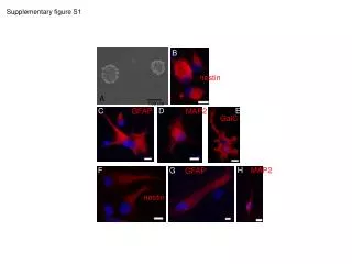
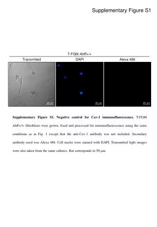
Supplementary Figure S1: T-FGM AhR +/+ fibroblasts as a negative control for Cav-1 immunofluorescence analysis. Fixed cells processed without anti-Cav-1 antibody, stained with Alexa 488 for nuclei and DAPI. Transmitted light images captured. Bar scale: 50mm.

E N D
Supplementary Figure S1 T-FGM AhR+/+ Transmitted DAPI Alexa 488 20 mm 20 mm 20 mm Supplementary Figure S1. Negative control for Cav-1 immunofluorescence. T-FGM AhR+/+ fibroblasts were grown, fixed and processed for immunofluorescence using the same conditions as in Fig. 1 except that the anti-Cav-1 antibody was not included. Secondary antibody used was Alexa 488. Cell nuclei were stained with DAPI. Transmitted light images were also taken from the same cultures. Bar corresponds to 50 mm.