
Chapter 7
Chapter 7. Cell Structure and Function (Aligned with 7.1 Intro Sheet and 7.2 Cell Structure Chart).

Chapter 7
E N D
Presentation Transcript
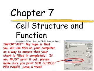
Chapter 7 Cell Structure and Function (Aligned with 7.1 Intro Sheet and 7.2 Cell Structure Chart) IMPORTANT: My hope is that you will use this on your computer as a way to ensure that your chart is filled in completely. If you MUST print it out, please make sure you print SIX SLIDES PER PAGE!! Save a tree!!
7-1 Life is Cellular A. Cells – basic units of structure and function in living things
B. Early scientists that led to the cell theory • Anton van Leeuwenhoek (1600s) – given credit for developing the 1st mini microscope, looked at pond water and made detailed drawings • Robert Hooke – coined the term “cell” when he looked at slices of cork and dead plant cells • Robert Brown (1833) – observed a dark structure near • the center of the cell (we now know this is the nucleus) • Matthias Schleiden (1838) – stated all plants are made • of cells • Theodore Schwann (1839) – stated all animals are made from cells • Rudolph Virchow (1855) - stated all cells come from the division of preexisting cells • Lorenz Oken – stated all new cells are the result of division of preexisting cells…VIRCHOW STOLE HIS THUNDER
C. Cell Theory • All living things are composed of cells. • Cells are the basic units of structure and function in living things. • All cells come from preexisting cells.
Exploring the Cell : Microscopes LM 1,000 TEM 2,800 SEM 2,000 Figure 4.1D Figure 4.1C Figure 4.1B The light microscope (LM): • Enables us to shape and structure of a cell • Magnify cells up to 1,000X • The electron microscope (EM): • Allows greater magnification and reveals cellular details • Magnify cells up to 2 mill X • Limits: no living specimens Transmission EM: (TEM) inside details Scanning EM: (SEM) surface structures * Lenses used to magnify image by focusing beams of light OR electrons • Magnification = image size * Resolution = clarity of detail
Why are cells so small? • A small cell allows a sufficient surface area to allow nutrients and wastes to cross per it’s volume. • (in other words: it can’t get too big for it’s own good) volume= 27,000 Ratio: .6 to 1 volume= 27,000 Ratio: .2 to 1 need as high a surface area to volume ratio as possible
What is in a Cell? ALL cells: * contain cytoplasm, cell membrane, and DNA * are either a prokaryotic or a eukaryotic
7-2 Cell Structure • Cellular Control Center • Nucleus, nucleolus, chromatin/chromosomes, nuclear membrane/pore • Organelles that Store, Clean Up and Support • Vacuoles, vesicles, lysosomes, cytoskeleton (microtubules, microfilaments), centrioles • Organelles that Build Proteins • Ribosomes, endoplasmic reticulum (smooth and rough), golgi apparatus • Organelles that Capture and Release Energy • Chloroplasts, mitochondria • Cellular Boundaries • Cell wall, cell membrane • Fluid Portion Outside of Nucleus (Sort of a boundary!) • cytoplasm
CELLULAR CONTROL CENTER: NUCLEUS • Function: • Information center of the cell • Contains DNA (chromatin vs. chromosomes) • Directs cell activities • 2 categories of organisms • Prokaryotes – organisms whose cells lack nuclei (i.e. bacteria) • Eukaryotes – organism whose cells contain nuclei
Nucleus Structure: • NUCLEOLUS – a small, darkened region in the nucleus that is made up of RNA and proteins, this is where ribosomes are made • CHROMOSOMES – large structures formed from DNA that contain the genetic info • CHROMATIN – uncondensed DNA found in non-dividing cells • NUCLEAR ENVELOPE – double membrane around the nucleus that contains pores, allows molecules to move in and out of the nucleus, and protects the nucleus • NUCLEAR PORES – allows passage of materials into or out of nucleus (RNA, ribosomes)
ORGANELLES THAT STORE, CLEAN UP, SUPPORT: VACUOLE • Structure: • Large, central structure in plants • Many, small, circular structures in animal cells • Filled with liquid • Function: • Storage of water, salts, proteins, carbohydrates, waste products • Pressure system for plants, prevents wilting • Special case: contractile vacuole - prevents excess water intake, leading to cell-bursting
ORGANELLES THAT STORE, CLEANUP, SUPPORT: VESICLE • Store and move materials between cell organelles and to/from cell surface
ORGANELLES THAT STORE, CLEAN UP, SUPPORT:LYSOSOME • Structure: • Small, circular structures • Found only in animal cells • Contain digestive enzymes • Function: • Digestion of: • Worn out organelles • Debris • Large ingested particles • Lysosomes are responsible for your hands not being webbed!!
ORGANELLES THAT STORE, CLEAN UP, SUPPORT: CYTOSKELETON • Structure: • Hollow tubes of proteins • Examples: cilia (cells lining tracea), flagella (sperm cells), centrioles • Function: • Framework • Provide cell with support, structure and shape • Movement (cilia, flagella) • Microfilaments – made of actin threads,allow movement of cytoplasm within the cell (cytoplasmic streaming) • Microtubules – hollow structure made of tubulin, maintain cell shape, make up cilia, flagella and centrioles
ORGANELLES THAT STORE, CLEAN UP, SUPPORT: CENTRIOLES • Organize cell division
ORGANELLES THAT STORE, CLEAN UP, SUPPORT:PLASTID • Structure: • Differ based on type of plastid (chloroplast is one example) • Found only in plants • Function: • Store food/starch • Store pigments (give color to fruits & veggies)
ORGANELLES THAT BUILD PROTEINS: RIBOSOMES • Structure: • Small (25 nm) ball-like structures • Found free-floating in cytoplasm or attached to rough endoplasmic reticulum • Composed of RNA and protein • Make peptide bonds between amino acidsprotein • Function: • Synthesis of proteins (where proteins are made)
ORGANELLES THAT BUILD PROTEINS: ENDOPLASMIC RETICULUM • Structure: • Network of flattened sacs • Can be rough (w/ ribosomes) or smooth (w/o) • Function: • Transport materials within or out of cell • Synthesis of macromolecules • Rough - proteins, lipids, carbs • Smooth - lipids
ORGANELLES THAT BUILD PROTEINS: GOLGI APPARATUS • Structure: • Flattened stacks of membranes • Vesicles attached to top and bottom • Function: • Collection, modification, packaging of proteins and other substances • Vesicles attach, deposit materials • GA modifies materials based on needs • Vesicles attach to membrane and distribute modified substances
ORGANELLES THAT CAPTURE/RELEASE ENERGY:CHLOROPLAST • Structure: • Double membrane • Elaborate structure inside • Function: • Another power station • Found in plant cells only • Conversion of light energy (sun) into chemical energy (glucose)
ORGANELLES THAT CAPTURE/RELEASE ENERGY:MITOCHONDRIA • Structure: • Double membrane • Cristae - inner folds, increase surface area • Outer membrane for protection of cell • Function: • “Powerhouse” of the cell • Able to self-replicate ( # in cells with high energy need) • Converts sugars into energy for cells
CELLULAR BOUNDRIES: CELL WALL • Only in plants, algae, and some bacteria • Lies outside the cell membrane • Function • Helps to protect and support the cell • Very porous (water, oxygen, carbon dioxide, etc. can pass through easily) • Gives rectangular shape to plant cells • Layers • 1st layer – contains pectin (gluey substance that helps hold the cells together) • 2nd layer – primary cell wall (made of cellulose) • 3rd layer (in woody stems) – secondary cell wall (composed of cellulose and lignin to make cellulose more rigid)
CELLULAR BOUNDRIES:CELL MEMBRANE – outer boundary • Structure: • Phospholipid bilayer • hydrophilic heads, hydrophobic tails • Contains lipids (bilayer), proteins (channels), and carbohydrate chains (identification cards) • Function: • Regulates what enters and leaves the cell • Semi-permeable membrane • Protection and support
CELLULAR BOUNDRIES:CYTOPLASM • Material between the cell membrane and the nucleus • Contains the organelles of the cell
QUESTIONS: • Describe the steps involved in the synthesis, packaging, and exporting of a protein from a cell. • What are the two major parts of the cell? • How do contractive vacuoles help maintain water balance? • What is the difference between rough and smooth ER? • Why is the cell membrane sometimes referred to as a fluid mosaic? What part of the cell membrane acts like a fluid? And what makes it like a mosaic? • How do the properties of lipids help explain the structure of the cell membrane? • Why do you think it’s important that cell membranes are selectively permeable?
QUESTIONS: • Describe the steps involved in the synthesis, packaging, and exporting of a protein from a cell. • Proteins assembled on ribosomes (if targeted for export to cm or to specialized locations w/in cell, complete their assembly on RER protein in vesicle Golgi apparatus (further modifies proteins before sorting and packaging them in membrane bound vesicles) vesicle final destination • What are the two major parts of the cell? • Cytoplasm with organelles, and nucleus • How do contractive vacuoles help maintain water balance? • Pump out excess water • What is the difference between rough and smooth ER? • Rough has ribosomes, smooth does not • Why is the cell membrane sometimes referred to as a fluid mosaic? What part of the cell membrane acts like a fluid? And what makes it like a mosaic? • It is made of many parts (like a mosaic) that can float around in the fluid phospholipid bilayer • How do the properties of lipids help explain the structure of the cell membrane? • Hydrophilic lipid heads are attracted to water, hydrophobic fatty acid tails turn away from water. A bilayer forms when heads turn outward towards water inside and outside a cell • Why do you think it’s important that cell membranes are selectively permeable? • Allows needed substances to enter and wastes to leave, while keeping molecules that are not needed out


