Download
1 / 43
430 likes | 716 Views
Welcome to Cell Biology!. it's alive!. Cell Theory. 2.1: Outline the Cell Theory 2.1.2: Discuss the Evidence for Cell Theory. Cell Theory. Living organisms are composed of cells Cells are the smallest unit of life

E N D
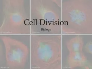
Welcome to Cell Biology! • it's alive!
Cell Theory • 2.1: Outline the Cell Theory • 2.1.2: Discuss the Evidence for Cell Theory
Cell Theory • Living organisms are composed of cells • Cells are the smallest unit of life • Cells come from pre-existing cells, by division (so that new cells cannot be constructed from non living substance)
The History of Cell Theory Let’s meet who discovered cells… Meet the Scientists...
1. All living things are made of cells… Digestive Tissue Goblet Cells http://www.olympusmicro.com/galleries/abramowitz/index.html 6.11.12
How do we know that all living things are made of cells? Because we can SEE them through microscopes….
When we we first start talking about ‘Cells’? Robert Hooke coined the term ‘cells’ since he felt that the space-filled chambers of dead cork resembled a monk’s empty cell…
Cell Theory: Robert Hooke Micrographia, 1665: . . . I could exceedingly plainly perceive it to be all perforated and porous, much like a Honey-comb, but that the pores of it were not regular. . . . these pores, or cells, . . . were indeed the first microscopical pores I ever saw, and perhaps, that were ever seen, for I had not met with any Writer or Person, that had made any mention of them before this. . .
The Father of Microscopy: Van Leeuwenhoek The first man to visualise single-celled animals (he called protistsanimacules): 1674 Microscopy bytes
Nothing smaller can survive independently If a cell is broken down into its individual components, its subunits cannot survive independently
3.All cells come from pre-existing cells: ‘Omnis Cellula e cellula ‘ • Disproving spontaneous generation • Pasteur
Limitations 1: There are many unicellular organisms • Mobile amoeba Primitive unicellular organisms carry out all of the functions of life • Prostista • Paramecium • Amoebae • Giant amoebae!
Limitations to cell theory: multinucleated cells (syncytiae) • Skeletal and cardiac muscle
Limitations to cell theory: multinucleated cells (syncytiae) Fungal hyphae (phase contrast microscopy)
Cell Biology: Magnification and Illumination Or…how to win a Nobel prize….
Cell Biology: Magnification and Illumination • Let's take a look at the secret life inside our cells...
IB Cell Theory 2.1.4 ‘Compare the relative sizes of molecules, cell membrane thickness, viruses, bacteria, organelles and cells, using the appropriate SI unit.’
How large are cells? Let's put things into perspective....
Is magnification all that matters?Magnification versus Resolution • Is there a limit to magnification? • Does magnification improve resolution? • Resolution of a microscope is its ability to separate small objects which are cose together • Resolution is determined by light/(electron) wavelength; the shorter the wavelength, the higher the resolution • Light microscope resolution is 0.2 μm • Electron microscope resolution is 1 ηm • Scanning Tunnel microscope resolution is 0.01 ηm length 0.01 ηm depth
Light microscope: Magnification Normal maximum magnifications of ocular and objective lenses are 10X and 100X respectively, giving overall maximal magnification of X 1000
How do microscopes work (I)? • Anatomy of vision
How do microscopes work (2)? • Objective lens (high powered magnifying glass) • Very short focal length (very close to the specimen) • Inverted image at high magnification • Second weak lens (eyepiece) produces a real image
Modern Illumination techniques used in light microscopy These techniques modify the light path to generate improved contrast: • Phase contrast micrcoscopy • Cross-polarised light microscopy • Dark field microscopy • Fluorescent microscopy
Phase contrast microscopy Phase shift in-vivo
Phase contrast microscopy • Improved contrast, allowing identification of structures in living cells • Allowed us to understand cell division • Won its inventor, Franz Zernike, the NOBEL PRIZE in 1951 • Nobel Prize link to Phase microscope: • Phase Nobel
Contrast Microscopy • Fluorescence contrast techniques • Immunofluorescence techniques • Here is a whole gallery of beautiful images: • Fluorescence Gallery • Cell fluorescence
Electron Microscopy! • Follows the same principles as light microscopy, but shines a beam of electrons rather than light particles • The lower ‘wavelength’ of the electron beam allows incredible resolution • Can visualise particles to the order of a few angstom (10-10m)
Transmission Electron Microscopy • Image gallery • Designed by Ernst Ruska (Heidelberg) in 1938 • He won the Nobel Prize just before his death, in 1986 • First electron microscope was built in Toronto in 1938
Scanning Electron Microscope • a tour of the scanning electron microscope
Scanning Tunneling Microscope • Sharing a Nobel Prize • Scanning tunneling microscopes
IB 2.1.5 Calculate the linear magnification of drawings and the actual size of specimens in images of known magnification
Magnification and scale bars • You will often need to calculate the actual size of a specimen/ component of a cell from a microscope image, or a photograph/micrograph • The first step is to ensure that all parts of your calculation have the same units!! Magnification = size of image actual size of specimen
Calculating size/ Magnification Graticules can be used to help estimate organelle/ sample size
On Monday, we will learn about: • What limits cell size? • Why and how do cells ‘specialise’? • What are stem cells and why are they controversial?