Joints and Muscle Tissue Histology
Explore different types of joints, synovial joint classifications, and muscle tissue components and histology. Discover the structural details and functions of various joint types. Delve into the components of muscle tissue and its histological features.

Joints and Muscle Tissue Histology
E N D
Presentation Transcript
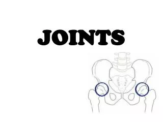
Joints Chapter 7 Bio160
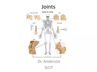
Types of Joints Fibrous – Sutures between skull bones, between teeth and jaw and between radius and ulna and tibia and fibula Cartilaginous – Epiphyseal plate, costal cartilage, between vertebrae and pubic symphysis
Types of Synovial Joints • Gliding – intercarpal and intertarsal, sternoclavicular, acromioclavicular, sternocostal, and vertebrocostal • Hinge – knee, elbow, ankle and interphalangeal • Pivot – atlanto-axial, radioulnar • Condyloid – wrist and metacarpophalangeal (2-5)
Types of Synovial Joints • Saddle – carpometacarpal in thumb • Ball and Socket – shoulder and hip
Types of Movements at Synovial Joints • Flexion and Extension • Rotation • Abduction and Adduction • Inversion and Eversion • Elevation and Depression • Supination and Pronation • Dorsiflexion and Plantar Flexion
Muscle Tissue Chapter8 Bio160
Connective Tissue Components • Entire muscle is wrapped in fibrous connective tissue which is continuous with tendons that insert skeletal muscle into bone • This outer connective tissue of muscle can also be called deep fascia (sheet of fibrous connective tissue) as compared to superficial fascia also known as subcutaneous tissue
Connective Tissue Components • Connective tissue holds muscle together as well as serving to transmit blood vessels and nerves to inner muscle cells • Connective tissue layers of skeletal muscle Epimysium - outside of entire muscle Perimysium - divides muscle cells into fascicles (bundles) Endomysium - covers individual muscle cells
Histology • Large → Small: fascicle → fiber → fibril → filament • Muscle consists of elongated cells called muscle fibers Sarco = fleshy • Sarcolemma - cell membrane
Histology • Transverse (T) tubule - tubular invagination of sarcolemma that surrounds each myofibril • Sarcoplasm – cytoplasm • Sarcoplasmic reticulum (SR) - smooth endoplasmic reticulum that stores Ca2+, has enlarged portions called cisternae that surround the transverse tubules
Histology • Myofibrils – cross section of muscle cell consists of small cylinders called myofibrils which may number several 100 to several 1000/cell (exercise increases myofibril production; lack of exercise decreases myofibrils (atrophy)) Each myofibril consists of myofilaments (protein)
Histology • thick myofilaments = myosin • thin myofilaments = actin, troponin, tropomyosin
Histology • thick myofilaments = myosin • thin myofilaments = actin, troponin, tropomyosin Each myofibril is surrounded by SR
Histology • Sarcomere - myofilaments don't extend entire length of muscle fiber; they are stacked into compartments called sarcomeres Sarcomeres are the functional unit of a skeletal muscle (contractile unit) Myofilament arrangement in sarcomeres results in alternating pattern of light and dark staining bands
Histology (6) Parts of sarcomere • A band - myosin + overlapping actin, dark staining band • I band - only actin, troponin, tropomyosin (2 I bands / sarcomere), light staining band • Z disc – through center of I band