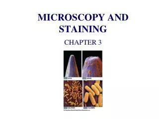
Microscopy and Staining
Microscopy and Staining. Unit of Measure. Bacteria are VERY small, that ’ s why this is micro biology The standard unit of measure in microbiology is the MICROMETER ( µm) A micrometer is 10 -6 or .000001M

Microscopy and Staining
E N D
Presentation Transcript
Unit of Measure • Bacteria are VERY small, that’s why this is micro biology • The standard unit of measure in microbiology is the MICROMETER (µm) • A micrometer is 10-6 or .000001M • To see something this small you need to use a microscope and also color (stain) the cells to see them
Microscope • Because bacteria are so small a microscope is the essential tool in microbiology • Light microscope uses visible light to observe bacteria
Resolution • Ability of the lens to distinguish fine detail • How close together can you distinguish two points as separate? • Because bacteria are so small good resolution is important • Resolution is DIRECTLY related to light in the following way: The SHORTER the wavelength of light the GREATER the resolution
More Resolution • To get the best resolution, use the SHORTEST wavelength of visible light that you can • Our microscopes use blue wavelength light to maximize resolution • Using blue light we can get a resolution of about .9 micrometers (µm)
Spectrum • Average wavelength of visible light is .55µm • Red light wavelength is .68µm, violet light is .42µm, blue light is .48µm • Which light is best to use? The one with shortest wavelength • Using a shorter wavelength of light in the blue range give better resolution
Smears and Staining • Bacteria must be stained (dyed) so they can be seen with the microscope • Before staining a smear must be made • A smear is just a film of bacteria on a glass slide • After the smear dries it is heat fixed, this • Kills the bacteria • Helps adhere the cells to the slide • Makes the cells more receptive to the dye
Stains • Stains are dyes • Stains carry either a positive charge (basic dyes) or a negative charge (acidic dyes) • Bacteria typically carry a slight negative charge on the cell surface so they attract a basic dye • Most of the stains used in the lab are basic dyes • A negative stain uses acidic dyes that do not stain the cell but rather the background
Simple Stain Uses only one basic dye Provides basic information about cell shape and arrangement Differential Stain Uses more than one dye These procedures react differently with different kinds of bacteria Helps distinguish between different kinds of bacteria Most common and important differential stain is the GRAM STAIN Staining Techniques
Gram Stain • Most important differential staining technique • Differentiates all bacteria based on cell wall composition • Bacteria are either Gram + and stain blue or Gram- and stain red • Gram stain is usually the first step in identifying an unknown bacteria
Acid-fast Stain • Differential stain • Identifies bacteria with MYCOLIC ACID in their cell walls • Very important human pathogens that can be identified with this stain is Mycobacterium tuberculosis • All members of genus Mycobacterium are acid-fast
Special stains • Negative stain • Acidic dye stains background, not cell • Used to determine cell shape and size • Spore stain • Used to identify bacteria that can form spores