Download
1 / 78
840 likes | 1.2k Views
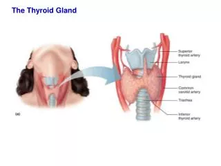
Thyroid gland 1 & 2. Dr Heyam Awad FRCPath. Thyroid gland. Thyroid hormones. Parafollicular cells. Thyroid diseases. Hyerthyroidism Hypothyroidism Thyroiditis Graves disease Diffuse nontoxic goiter and multinodular goiter neoplasms. THYROTOXICOSIS

E N D
Thyroid gland 1 & 2 DrHeyamAwad FRCPath
Thyroid diseases • Hyerthyroidism • Hypothyroidism • Thyroiditis • Graves disease • Diffuse nontoxic goiter and multinodular goiter • neoplasms
THYROTOXICOSIS Associated with hyperthyroidism (Thyroid hyperfunction): 1. Primary a. Diffuse toxic hyperplasia (Graves disease) b. Hyperfunctioning (Toxic) multinodulargoiter) • Hyperfunctioning (toxic ) adenoma • Secondary -- TSH-secreting pituitary adenoma (rare) Thyrotoxicosisnot associated with hyperthyroidism is less common • Excessive release of pre-formed hormone in thyroiditis • Ectopic secretion of thyroid hormones.
Clinical manifestations of thyrotoxicosis a. Constitutional symptoms : warm flushed skin, heat intolerance and excessive sweating - Weight loss despite increased appetite. b. Malabsorption, and diarrhea. c. Tachycardia and elderly patients may develop heart failure due to aggravation of pre-existing heart disease d. Nervousness, tremor, and irritability
e. A wide, staring gaze and lid lag because of sympathetic overstimulation of the levatorpalpebraesuperioris Note: True thyroid ophthalmopathy associated with proptosisis a feature seen only in Graves disease. f. 50% develop proximal muscle weakness (thyroid myopathy). g. Thyroid storm : Designates the abrupt onset of severe hyperthyroidism, and this condition occurs most commonly in individuals with Graves disease and it is a medical emergency because significant numbers of untreated patients die of cardiac arrhythmias
Lab tests • The measurement of serum TSH is the most useful single screening test for hyperthyroidism, because TSH levels are decreased even at the earliest stages, when the disease may still be subclinical
- Once the diagnosis of thyrotoxicosis has been confirmed measurement of radioactive iodine uptake by the thyroid gland often is valuable in determining the etiology For example, such scans may show : a. Diffusely increased (whole-gland) uptake in Graves disease, b. Increased uptake in a solitary nodule in toxic adenoma c. Or decreased uptake in thyroiditis.
HYPOTHYROIDISM : Primary causes a. - Worldwide, the most common cause of hypothyroidism is dietary deficiency of iodine. b. In most developed countries, autoimmune diseases predominate such as Hashimoto thyroiditis c. Genetic defects such as Thyroid dysgenesis or Congenital biosynthetic defect (dyshormogenticgoiter). Secondary causes: Pituitary or hypothalamic disorder
hypothyroidism • Cretinism • myxedema
Cretinism :Refers to hypothyroidism developing in infancy or early childhood • Endemic cretinism: in dietary iodine deficiency is endemic, including mountainous areas ( the Himalayas ) 2. Sporadic cretinism. Caused by enzyme defects that interfere with thyroid hormone synthesis
Clinical features of cretinism include: - Impaired development of skeletal system- short stature, - Coarse facial features, protruding tongue, umbilical hernia. • Central nervous system, with mental retardation
Myxedema. or Gull syndrome : • Hypothyroidism in older children and adults and characterized by: a. Patients are cold intolerant, and often obese. b. Generalized apathy and mental sluggishness that in the early stages of disease may mimic depression c. Broadening and coarsening of facial features
d. Enlargement of the tongue, and deepening of the voice. e. Bowel motility is decreased, resulting in constipation. f. Pericardial effusions are common; in later stages, the heart is enlarged, and heart failure may supervene. • Mucopolysaccharide-rich edematous fluid accumulates in skin, subcutaneous tissue, and number of visceral sites
Lab tests Serum TSH is the most sensitive screening test . a. The serum TSH is increased in primary hypothyroidism b. The TSH is not increased in persons with hypothyroidism caused by primary hypothalamic or pituitary disease. c. Serum T4 is decreased hypothyroidism of any origin.
Thyroiditis Chronic Lymphocytic (Hashimoto) Thyroiditis - Is the most common cause of hypothyroidism in areas of the world where iodine levels are sufficient. - It is characterized by gradual thyroid failure secondary to autoimmune destruction of the thyroid gland - It is most prevalent between the ages of 45 and 65 years and is more common in women than in men - It can occur in children and is a major cause of non-endemic goiter in children
PATHOGENESIS :- Caused by breakdown in self-tolerance to thyroid antigens - Circulating autoantibodies against thyroid antigens are- present in the vast majority of patients - Multiple immunologic mechanisms may contribute to thyroid damage , • Cytokine-mediated cell death: Excessive T cell activation leads to the production of inflammatory cytokines such as IFN-γ in the thyroid with resultant recruitment and activation of macrophages and damage to follicles . • Binding of anti-thyroid antibodies (antithyroglobulin, and antithyroid peroxidase antibodies), followed by antibody- dependent cell-mediated cytotoxicity • T cell mediated cytotoxicity.
. HASHIMOTO - A significant genetic component is supported by the a. Concordance of disease in 40% of monozygotic twins, b. the presence of circulating antithyroid antibodies in 50% of asymptomatic siblings of affected patients .
Gross : - Diffuse and symmetric enlargement of the thyroid but localized enlargement may be seen in some cases to raise suspicion for neoplasm Microscopic examination reveals 1. Infiltration by small lymphocytes, plasma cells, and well-developed germinal centers 2. The thyroid follicles are atrophic 2. Some follicles are lined by epithelial cells with abundant eosinophilic, cytoplasm, termedHürthle cells and these Hurthle cells have numerous mitochondria
- Less commonly, the thyroid is small due to extensive fibrosis (fibrosing variant) but unlike Reidelthyroiditis, the fibrosis does not extend beyond the capsule of the gland. Clinically , • Painless thyroid enlargement associated with some degree of hypothyroidism, • - In the usual clinical course, hypothyroidism develops gradually.; however, it may be preceded by transient thyrotoxicosis due to disruption of thyroid follicles ,and secondary release of thyroid hormones (hashitoxicosis).
- Patients with Hashimoto thyroiditis often : 1. Have other autoimmune diseases 2. .Are at increased risk for the development of B cell non-Hodgkin lymphomas within the thyroid gland. Note: - The relationship between Hashimoto disease and thyroid epithelial cancers remains controversial, with some morphologic and molecular studies suggesting a predisposition to papillary carcinomas
Subacute Granulomatous (de Quervain) Thyroiditis - Is much less common than Hashimoto disease - Is most common between the ages of 30 and 50 and, - More frequently in women than in men. - Is believed to be caused by a viral infection and a majority of patients have a history of an upper respiratory infection just before the onset of thyroiditis. Gross- The gland has intact capsule, and may be unilaterally or bilaterally enlarged.
Histologic examination reveals 1. Disruption of thyroid follicles, with extravasation of colloid leading to a neutrophilic infiltrate, which is replaced by lymphocytes, plasma cells, and macrophages. 2. The extravasated colloid provokes a granulomatous reaction with giant cells that contain fragments of colloid. 3. Healing occurs by resolution of inflammation and fibrosis. Clinical Features : -Acute onset characterized by neck pain ( with swallowing) - Fever, malaise, and variable enlargement of the thyroid. - Transient hyperthyroidism may occur as a result of disruption of follicles and release of excessive hormones. - The leukocyte count is increased.
- With progression of disease and gland destruction, a transient hypothyroid phase may ensue. - The condition typically is self-limited, with most patients returning to a euthyroid state within 6 to 8 weeks
SubacuteLymphocytic Thyroiditis : - Also is known as silent or painless thyroiditis; - And in a subset of patients the onset of disease follows - pregnancy (postpartum thyroiditis). - Most likely to be autoimmune because circulating antithyroid antibodies are found in a majority of patients - It mostly affects middle-aged women, who present with a- painless neck mass or features of thyrotoxicosis
Riedel thyroiditis,: A rare disorder of unknown etiology, - Characterized by extensive fibrosis involving the thyroid and contiguous structures simulating a thyroid neoplasm • May be associated with idiopathic fibrosis in other parts of the body, such as the retroperitoneum - The presence of circulating antithyroid antibodies in most patients suggests an autoimmune etiology
GRAVES DISEASE The most common cause of endogenous hyperthyroidism with a peak incidence in women between the ages of 20 and 40. Triad of manifestations: A. Thyrotoxicosis,. All patients B. Localized, infiltrative dermopathy( pretibial myxedema), minority of cases and involves the skin overlying the shins, and manifests as scaly thickening C. Infiltrative ophthalmopathywith resultant exophthalmos in 40% of patients
Exophthalmos is the result of increased volume of the retro-orbital connective tissues by • Marked infiltration of T cells with inflammatory edema • Accumulation of glycosaminoglycans • Increased numbers ofadipocytes (fatty infiltration). • These changes displace the eyeball forward, potentially interfering with the function of the extraocular muscles • Exophthalmos may persist after successful treatment of the thyrotoxicosis, and may result in corneal injury.
PATHOGENESIS :- Genetic factors are important in the causation of Graves disease, the incidence is increased in relatives of affected patients, and the concordance rate in monozygotic twins is 60%. - A genetic susceptibility is associated with the presence of HLA-DR3, - it is characterized by a breakdown in self-tolerance to thyroid autoantigens, and is the production of multiple autoantibodies
Autoantibodies in GRAVES : 1. Thyroid-stimulating immunoglobulin: • An IgG antibody binds to the TSH receptor and mimics the action of TSH, with resultant increased hormones 2. Thyroid growth-stimulating immunoglobulins: - Directed against the TSH receptor, and have been implicated in the proliferation of follicular epithelium 3. TSH-binding inhibitor immunoglobulins: - Prevent TSH from binding to its receptor on thyroid cells and in so doing may actually inhibit thyroid cell function, a finding explains why some patients with Graves spontaneously develop episodes of hypothyroidism.
Note: The coexistence of stimulating and inhibiting immunoglobulins in the serum of the same patient may explain why some patients with Graves disease spontaneously develop episodes of hypothyroidism .Gross: Symmetrical enlargement of the thyroid gland with intact capsule,
On microscopic examination, a. The follicular cells in untreated cases are tall, and more crowded and may result in formation of small papillae b. Lymphoid infiltrates, consisting predominantly of T cells, with few B cells and plasma cells are present throughout the interstitium; with formation of germinal centers Laboratory findings and radiologic findings - Elevated serum free T4 and T3 and depressed serum TSH - Because of ongoing stimulation of the thyroid follicles radioactive iodine uptake is increased, and radioiodine scans show a diffuse uptake of iodine.
DIFFUSE AND MULTINODULAR GOITER - Enlargement of the thyroid, or goiter, is the most common manifestation of thyroid disease Mechanism : - The goiters reflect impaired synthesis of thyroid hormone often caused by dietary iodine deficiency and this leads to to a compensatory rise in the serum TSH, which in turn causes hyperplasia of the follicular cells and, ultimately, gross enlargement of the thyroid gland .,
Goiters can be endemic or sporadic. I. Endemic goiter :Occurs in geographic areas where the soil, water, and food supply contain little iodine. - The term endemic is used when goiters are present in more than 10% of the population in a given region. - Such conditions are common in mountainous areas of the world, including the Himalayas and the Andes but with increasing availability of iodine supplementation, the frequency and severity of endemic goiter have declined
II. Sporadic goiter : Less common than endemic goiter. - The condition is more common in females than in males, with a peak incidence in puberty or young adulthood, when there is an increased physiologic demand for T4. - It may be caused by several conditions, including the: a. Ingestion of substances that interfere with thyroid hormone synthesis , such as excessive calcium and vegetables such as cabbage, cauliflower, sprouts, . b. Hereditary enzymatic defects that interfere with thyroid hormone synthesis (dyshormonogenetic goiter). -In most cases, the cause of sporadic goiter is not apparent.
MORPHOLOGY : - Initially, the gland is diffusely and symmetrically enlarged (diffuse goiter) but later on it becomes multinodular goiter. On microscopic examination, a. The follicular epithelium may be hyperplastic in the early stages of disease or flattened and cuboidal during periods of involution. b. Colloid is abundant in the latter periods (colloid goiter). c. With time, recurrent episodes of hyperplasia and involution produce amore irregular enlargement of thee thyroid, termed multinodular goiter and virtually all long-standing diffuse goiters convert into multinodular goiters.
- Multinodular goiters cause multilobulated, asymmetrically enlarged glands which attain massive size and old lesions often show fibrosis, hemorrhage, calcification - Multinodular goiters typically are hormonally silent, - 10% of patients can manifest with thyrotoxicosis due to the development of autonomous nodules producing hormone independent of TSH stimulation and this condition, called toxicmultinodular goiter or Plummer syndrome
Clinical Features : a. The dominant features are mass effects of the goiter b. may cause airway obstruction, dysphagia, and compression of large vessels in the neck and upper thorax c. The incidence of malignancy in long-standing multinodular goiters is low (less than 5%) but not zero and concern for malignancy arises with goiters that demonstrate sudden changes in size or associated symptoms ( hoarseness)
Thyroid tumors : -present as solitary nodules. - themajority of solitary nodules of the thyroid prove to be benign : a. Follicular adenomas b. A dominant nodule in multinodular goiter • Simple cysts or foci of thyroiditis - Carcinomas of the thyroid, are uncommon, accounting for much less than 10% of solitary thyroid nodules.