The Immune System
The Immune System . Chapter 21. Immune System. functional system rather than organ system Hematopoetic Vasculature Lymphatic. Fig 21.1. Innate vs. Adaptive Immune System – Introduction. Innate: structural defenses; responds to nonspecific foreign substances

The Immune System
E N D
Presentation Transcript
The Immune System Chapter 21
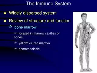
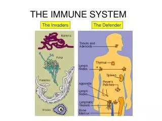
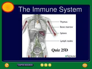
Immune System • functional system rather than organ system • Hematopoetic • Vasculature • Lymphatic Fig 21.1
Innate vs. Adaptive Immune System – Introduction • Innate: structural defenses; responds to nonspecific foreign substances • First line: external surface epithelium & membranes • Second line: inflammatory processes – antimicrobial proteins, phagocytes, etc. Fig 21.1
Innate vs. Adaptive Immune System – Introduction • Adaptive: responds to specific foreign substances • Innate & adaptive mechanisms work together Fig 21.1
Innate, Surface Defenses • Skin • physical barrier to microbes • Keratin resistant to most bacterial enzymes & toxins • secretions are acidic pH 3-5 • Mucosa • physical barrier & produces a variety of protective chemicals • Gastric mucosa • very acidic & produces proteolytic enzymes • Saliva & lacrimal fluid contain lysozyme • Mucous • traps bacteria & moves them away from epithelial surface
Innate, Internal Defenses • Based on recognition of surface carbohydrates (glycocalyx) • Glycocalyx is recognized as “self” or “non-self” Figure 3.3
Innate, Internal Defenses • Phagocytes • Macrophages: derived from monocytes • Free Macrophages: roam through tissues • Fixed Macrophages: Kupffer cells (liver) & microglia (brain) • Ingest cellular debris, foreign material, bacteria, fungi • Neutrophils: ingest pathogens • Eosinophils: weakly phagocytic of pathogens. Attack parasites (degranulation) • Mast Cells: phagocytic of various bacteria
Innate, Internal Defenses • Phagocytic mechanisms: • Adherence: cell binds to invader • Aided by opsonization (a chemical process that enhances binding via complement & antibodies) • Ingestion: formation of phagolysosomes • Respiratory Bursts: merge phagosome with lysosome & flood phagolysosome with free radicals (macrophage) • Defensins: proteins that crystallize out of solution & pierce pathogen membranes (neutrophils)
Mechanism of Phagocytosis Figure 21.2
Innate, Internal Defenses • Natural Killer Cells: • Small population of large granular lymphocytes • Non specific for “non-self” • Not phagocytic: attack is by release of perforins that perforate the target cell plasma membrane. • Shortly after perforation the target nucleus disintegrates. • Release chemicals that enhance the inflammatory response
Innate, Internal Defenses: Inflammation • tissue response to injury • Triggered by injury – trauma, heat, chemical irritation, infection, etc. • Beneficial effects • Prevents spread of injury • Disposes of cellular debris & pathogens • Promotes repair
Innate, Internal Defenses: Inflammation • cardinal signs of inflammation • Redness • Heat • Swelling • Pain • (functional impairment Rigor) • Weapons of the Spanish Inquisition
Innate, Internal Defenses: Inflammation • Inflammatory response: signs are associated with vasodilation & increased vascular permeability • Dilation: redness, heat • Permeability: edema, (increased pressure) pain • Pain also associated with bacterial toxins & some mediators (kinins, PGs)
Innate, Internal Defenses: Inflammatory Response • Mechanisms causing vasodilation & vascular permeability • Injured cells release inflammatory mediators • Histamines • Kinins • Prostaglandins • Complement • Cytokines (also activated by receptors on macrophages in response to microbial glycocalyx)
Innate, Internal Defenses: Inflammatory Response • Edema • Dilutes harmful substances • Provides nutrients (& O2) for repair • Enhances entry of clotting protein • Epithelial breaches also stimulate b-defensin release from epithelial cells
Events in Inflammation Figure 21.3
Innate, Internal Defenses: Inflammatory Response • Phagocyte mobilization: infiltration of damaged area by neutrophils & macrophages
Innate, Internal Defenses: Inflammatory Response • Leukocytosis: leukocytosis inducing factors released by injured cells promote rapid release of WBCs from marrow • Margination: increased vascular permeability causes decreased fluid in vessels; blood flow slows & neutrophils are able to move to vessel margins. Here endothelial markers (CAMs) allow neutrophils to cling to vessel walls (pavementing).
Innate, Internal Defenses: Inflammatory Response • Diapedesis: neutrophils migrate through capillary walls • Chemotaxis – inflammatory chemicals attract neutrophils to move up the chemical concentration gradient (neutrophils respond first) • As the process continues, monocytes diapedes into the area & become macrophages. With chronic inflammation, macrophages predominate
Inflammatory Response:Phagocytic Mobilization Figure 21.4
Innate, Internal Defenses: Inflammatory Response • Macrophages clean up cellular debris & pathogens • If pathogens were associated with the injury, activation of the complement cascade occurs & elements of adaptive immunity join the process
Innate, Internal Defenses • Viral replication – (viruses lack metabolic processes) Viruses release nucleic acid (RNA or DNA) into cytoplasm. The information on the nucleic acid is incorporated into the cell’s DNA. Normal cellular mechanisms then produce viral structural components. Multiple new viral particles are produced & released from the cell (sometimes killing the cell)
Innate, Internal Defenses • Antiviral proteins: interferon & complement • Interferon: some cells produce & release interferons (IFNs) when invaded by virus • Released interferons stimulate nearby cells to produce proteins (PKR) that interfere with viral replication by disrupting protein synthesis & the ribosome • Not virus specific.
Interferon (IFN) Figure 21.5
Innate, Internal Defenses • Complement – a group of plasma proteins (20) that are activated in the presence of foreign substances • Complement activation enhances & amplifies inflammation • Bacteria & some other cell types are lysed by complement activation • Complement activation enhances both innate & adaptive defenses
Innate, Internal Defenses • Complement activation pathways • Classical pathway: requires antibodies • Antibodies bind to target (antigen) • Complement protein C1 binds to the antibody-antigen complex (complement fixation) • Alternative pathway: complement factors interact with microorganism glycocalyx • Both pathways lead to a cascade of protein activation, leading to activation of C3
Innate, Internal Defenses • C3 is the start of the; Final Common Pathway • C3 cleaves to form C3a & C3b • C3a (& C5a) enhance inflammation by increasing histamine release, increasing vascular permeability & stimulating chemotaxis • C3b coats bacterial membrane supplying adhesion points (opsonization) • C3b initiates the cascade forming the membrane attack complex (MAC) • The MAC forms a hole in the cell membrane & enhances Ca2+ influx cell lysis
Innate, Internal Defenses; Complement Figure 21.6
Innate, Internal Defenses • C-reactive proteins (CRP) produced by the liver in response to inflammatory molecules can activate the classical pathway by binding to membrane & activating C1. Also participates in opsonization. • Fever – a systemic response to infection. Leukocytes & macrophages release pyrogens that raise the hypothalamic “set point” for temperature
ADAPTIVE DEFENSES • ADAPTIVE DEFENSES • Innate & adaptive mechanisms work together in a cohesive fashion
Adaptive Defenses: Characteristics • Specificity: directed at specific targets • Systemic: not restricted to initial site of infection / invasion • Memory: after initial exposure & activation, a more rapid & more vigorous response is made to subsequent exposures to pathogens • (secondary response)
Adaptive Defenses: Components • Humoral Immunity: (antibody mediated immunity) provided by antibodies floating free in body fluids • Cell mediated immunity: • lymphocytes directly attack specific invaders by lysis or indirect attack by initiating inflammation and/or activating other lymphocytes & macrophages
Adaptive, Humoral Immunity • Antigen = any substance that can mobilize the immune system & provoke an immune response* *Humoral and/or cell mediated
Adaptive, Humoral Immunity • Complete antigens (proteins, nucleic acids, lipids, polysaccharides): • Immunogenicity: the ability to stimulate specific lymphocytes & specific antibodies • Reactivity: the ability to react with activated lymphocytes & antibodies • Hapten (an incomplete antigen): a smaller molecule that is not immunogenic until attached to proteins
Adaptive, Humoral Immunity • Antigenic determinants: sites on an antigenic molecule that are immunogenic • Epitope • Major Histocompatibility Complex (MHC): cell surface glycoproteins associated with self recognition Figure 21.7
Adaptive Immune System: Cells • Lymphocytes • T-cells • B-cells • Antigen Presenting Cells (APCs)
Adaptive Immune System: Cells • Lymphocytes: initially uncommitted • T-cells: are sorted in the Thymus • Positive selection: recognize MHC survive • Negative selection: react against to self-antigens on MHC killed • 2% of initial T-cell precursors • T-cells manage the immune response • B-cells: are sorted in the marrow by an incompletely understood process Figure 21.9
Adaptive Immune System: Cells • Immunocompetence: as T- or B-cells mature they become immunocompetent, they display receptors on their cell membrane for a specific antigen. • All of the receptors on one cell are identical; immunity depends upon genetic coding for appropriate receptors.
Adaptive Immune System: Cells • Antigen Presenting Cells (APCs) • APCs ingest foreign material, then present antigenic fragments on their cell surface where they are recognized by T-cells • T-cells: respond to antigen only if it is displayed on plasma membrane. • APCs: Macrophages & B lymphocytes • Interactions between APCs & lymphocytes & lymphocyte-lymphocyte interactions are critical to immune response
Adaptive, Humoral response • Humoral response (clonal selection) • B-cells: Antigen challenge to naïve immunocompetent B-cell • Antigen binds to B-cell receptors & form cross-links between receptors • Cross linked antigen-receptor complex undergoes endocytosis; B-cell presents to T-cell
Humoral Immunity • Active humoral immunity: • B-cells encounter & respond to antigen to produce an antibody • Passive humoral immunity: • Introduced “non-native” antibody
Active Humoral Immunity • Naturally acquired: natural exposure to antigen (i.e. infection) • Artificially acquired: vaccines; dead/attenuated or fragmented pathogen injected to elicit an immune response • Bestow immunity without disease; primary response • Booster shots (secondary response); intensify response • Shortcomings – adverse reactions & the immunity is less durable (poor memory) & has less cell mediated component
Passive Humoral Immunity • Natural: maternal antibody crosses the placental barrier conferring temporary immunity to the baby (degrades after a few months) • Artificial: antibodies harvested from an outside source given by injection protect from immediate threat but no memory is formed (antitoxins, antivenins , gamma globulin, etc.)
Antibodies • A.K.A Immunoglobulins & gamma globulins • Structure • variable • hypervariable • constant Figure 21.13a
Antibodies • Constant (C) region defines antibody class • determines chemical & cellular interactions • determines how class functions to eliminate antigens
Antibody Classes • Antibody Classes: IgM, IgG, IgA, IgD, IgE (Ig = immunoglobulin)
Antibody Classes • IgG: the most abundant circulating Ig. The dominant circulating Ig of the primary & the secondary response. Crosses the placenta. Complement binding (Monomer). • IgA: the Ig of secretions. Helps prevent antigen penetration of membranes (Dimer). • IgD: the Ig of B-cell activation. Found on B-cell surface (Monomer).
Antibody Classes • IgM: occurs as a monomer & a pentamer • Occurs on the B-cell surface (Monomer). • The Ig of early primary plasma cell response, circulating antibody; a potent agglutinator. Complement binding (Pentamer).