Download
1 / 72
780 likes | 1.7k Views
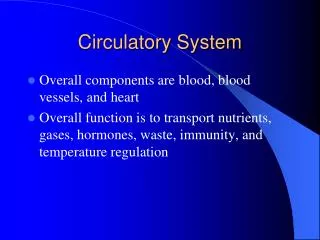
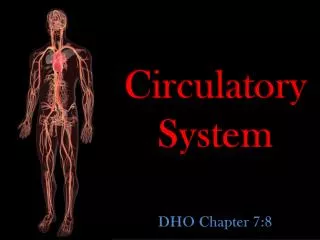
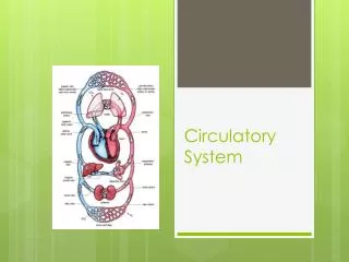
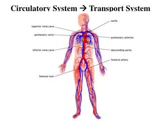
Circulatory System Transport System. We have two Circulatory Systems. 1 . Cardiovascular System. 2 . Lymphatic System. Thoracic Cavity Heart w/in mediastinum. 4 valves prevent backflow & blood moves in 1 direction. Aortic Semilunar Lt ventricle Aorta. Pulmonary Semilunar

E N D
We have two Circulatory Systems 1. Cardiovascular System 2. Lymphatic System
Thoracic Cavity Heart w/in mediastinum
4 valves prevent backflow & blood moves in 1 direction Aortic Semilunar Lt ventricle Aorta Pulmonary Semilunar Rt ventricle Pulmonary Artery Bicuspid/mitral A/V valve (2 flaps) Tricuspid A/V valve (3 flaps) Chordae Tendinae
Right DeO2 Left O2
Our Heart is like 2 Hearts in One • The Left Heart • Left Atrium • Left Ventricle • Pumps O2 Blood • to Body • The Right Heart • Right Atrium • Right Ventricle • Receives DeO2blood • from body
Right Left start start
Systemic Circulation Movement of blood from Lt Ventricle body includes hepatic portal circulation of liver & renal circulation of kidneys
Coronary Circulation To myocardium Rt coronary artery Rt coronary vein Lt coronary artery Lt coronary vein
Angina Pectoris: Severe chest pain when myocardium deprived of adequate O2. Coronary artery no longer supplies enough O2 to heart muscle. Veins harvested for bypass (prevent graph vs host rejection). Pg 259 fig 11-4
Heart Attack Myocardial Infarction Flow to a section of heart muscle becomes blocked. If not restored quickly, section of heart muscle damaged from lack of oxygen & begins to die. Damaged heart muscle loses its ability to contract Remaining heart muscle must compensate for weakened area.
Deflated balloon catheter inserted into narrowed coronary artery. Balloon is inflated, compressing the plaque & restoring size of artery. Relieves chest pain caused by reduced blood flow to the heart. Minimize damage to the heart muscle during a heart attack. This damage occurs when blood flow is totally cut off to an area of the heart. Stents: tiny mesh tube that is inserted in narrowed area to keep it open. Some coated with medication to help prevent the artery from closing again.
AbioCor artificial heart 2 pounds life expectancy 30 days – 6months Can extend life for those awaiting heart transplant donor
Heart Conduction Heart contracts in absence in external stimuli 4 structures embedded in heart wall generate & conduct electrical impulses through heart muscle cause atria & then ventricles to contract
Sinoatrial Node (SA node) Pacemaker in Rt Atrium wall Impulse starts & spreads in all directions through both atria contraction Impulse reaches Atrioventricular Node (AV node) between Rt atrium & Rt. ventricle Relayed to Bundle of His (AV bundle) Rt & Lt branches along septum and Purkinje fibers 0ff 2 AV bundles To ventricles contraction Disease can damage conduction system
Artificial Pacemaker Electrical device causes heart contractions maintains adequate blood flow 2 components to pacemaker *pulse generator, computer chip (brains) & battery *wires (leads) carry electrical signals to & from heart. Pulse generator under skin sends electrical signal to heart. 2 leads inserted through an arm vein are attached to the heart's Rt atrium or Rt ventricle or both.
Electrocardiogram (ECG or EKG) Heart conduction generates electrical currents picked up from body surface Graphic record of heart’s electrical activity 3 waves: deflection represents electrical activity associated with contraction & relaxation of atria & ventricles Damage to cardiac muscle affects conduction system distinct changes in ECG diagnosis & treatment
Depolarization: electrical activity w/ contraction of heart muscle Repolarization:relaxation of heart muscle P wave: atrial depolarization (contraction) QRS complex: ventricular depolarization (contraction) T wave: ventricular repolarization (relaxation) *Atrial repolarization wave masked by QRS complex
Vessel Structure Arteries & Veins: 3 layers Tunica Externa outermost Tunica Media middle Thicker in arteries vs veins maintain BP for blood distribution to body Smooth muscle: Autonomic NS control Tunica Interna innermost (fibrous, endothelial tissue) CNS (brain/spinal cord) NS sensory PNS somatic (skeletal muscle/effecters) motor autonomic (sm & cardiac muscle & glands/effecters) ↑sympathetic ↓parasympathetic adrenaline ↑ SA node impulses acetylcholine ↓ SA Node impulses
Arteries • Carry blood Away from the Heart • Are DEEP, near Bones for protection
Blood Vessels • Arteries: carry blood away from heart • Arterioles capillaries • O2 (except for pulmonary arteries) • under pressure • pre-capillary sphincters regulate blood • flow into capillaries (deeper than veins) • Blocked carotid artery stroke • Prevent with Endarterectomy
Arteries have a thick (Invol.) muscular wall Blood is moving very fast, under high pressure Smaller Arteries branch into smaller Arterioles Many arterioles have sphincters
Veins: carry blood to heart are superficial • Capillaries venules veins • DeO2 (except for pulmonary veins) • little to no pressure • valves prevent backflow • muscle contractions keep blood moving • in veins towards heart • (more superficial than arteries)
Varicose Veins Faulty valves in the veins blood pools distends veins Treatment Sclerotherapy: Injection causes vein to seal shut scars fades Laser surgery: Direct & Accurate Sends strong bursts of light onto vein fades Surgical Ligation and Stripping: Veins tied & removed Deeper veins take over circulation for treated veins
Capillaries Connect Artery to Vein Capillary bed diffusion gas/nutrientexchange O2 DeO2 venule arteriole
Where does Blood do All its Work? WHY??? Walls of Arteries & Veins are too thick and Blood is moving much too fast to do its work!
Blood Pressure forces some of the Plasma minus large blood proteins to leak out of Capillary (Filtration) into the Tissues Exchange of ALL Nutrients, Gases, & Wastes occurs here Too much Tissue Fluid = Edema