Download

1 / 66
690 likes | 1.09k Views
12. Lecture Note PowerPoint Presentation. The Integument. LEARNING OUTCOME 1. Describe normal skin changes associated with aging. . Normal Structure and Function of the Skin. Skin consists of 15–20% of the total body weight Epidermis

E N D
12 Lecture Note PowerPoint Presentation The Integument
LEARNING OUTCOME 1 Describe normal skin changes associated with aging.
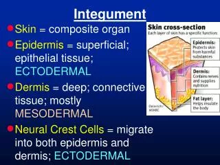
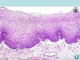
Normal Structure and Function of the Skin • Skin consists of 15–20% of the total body weight • Epidermis • Consists of five continually regenerating and shedding layers • Dermis
Normal Structure and Function of the Skin • Subcutaneous layers • A specialized connective tissue attached to muscles • Contains blood vessels, lymphatic channels, hair follicles, and sweat glands
Normal Structure and Function of the Skin • Accessory structures • Hair • Nails • Glands • Sebaceous glands • Apocrine sweat glands
Normal Structure and Function of the Skin • Function • Protection • Regulation of immune functions • Thermoregulation • Vitamin synthesis • Sensory receptor for CNS
Skin Changes Associated with Aging • Intrinsic factors • Genetic makeup and the normal aging process • Extrinsic factors • UV lighting • Smoking • Environmental pollutants
Figure 12-2Normal changes of aging in the integumentary system.
Skin Changes Associated with Aging • Epidermal changes • Thinning • Reduced moisture leading to a dry, rough appearance • Mitosis slows after age 50 by 30% • Increased healing time • Increased risk of infection
Skin Changes Associated with Aging • Epidermal changes • Rete ridges flatten: in the dermal layer, less collagen is being produced. The elastin fibers also wear out. Such factors will cause the skin to sag and wrinkle. The reteridges, meanwhile, will flatten out. This will cause the skin to be fragile. • Increased risk of skin breakdown • Reduced melanocytes • Paler complexion • Increased risk of UV damage
Skin Changes Associated with Aging • Epidermal changes • Scattered pigmented areas • Nevi (skin moles) • Age spots • Liver spots • Increased number and size of freckles (clusters of concentrated melanin)
Skin Changes Associated with Aging • Dermal changes • Decreased thickness and function begin in 3rd decade of life • Elastin decreases in quality • Wrinkling and sagging • Collagen less organized • Loss of turgor
Skin Changes Associated with Aging • Dermal changes • Reduced vascularity • Paler complexion • Capillaries thin and are easily damaged • Senile purpura Easy skin bruising in older people • Reduced touch and pressure sensations
Skin Changes Associated with Aging • Subcutaneous layer • Tissue thins in the face, neck, hands, and lower legs • Visible veins in exposed areas • Hypertrophy of tissue in certain body areas • Increased body fat • Increased body fat in abdomen and thighs
Hair Changes with Aging • Reduced number of functioning melanocytes • Replacement of pigmented strands of hair with nonpigmented hair • Hormone levels decline • Loss of hair in pubic and axillary areas • Growth of facial hair in women • Growth of nasal and ear hair in men • Increased baldness
Nail Changing with Aging • Color changes • Dull • Yellowing or grayness • Slowed growth • Thicker nails prone to splitting • Longitudinal striations • Related to damage at the nail matrix (the ROOT of the nail)
Nail Changing with Aging • Longitudinal pigmented bands • Single or multiple brown or black bands on thumb and index finger • Frequently seen in African-Americans over age 20 • Increased visibility in the older adult
Glandular Changes with Aging • Eccrine or sweat glands • Decreased number; decreased ability to regulate body temperature • Sebaceous glands • Increased size; decreased activity; increased water evaporation causes cracked, dry skin
LEARNING OUTCOME 2 Identify risk factors related to common skin problems of older adults.
“The Sun Never Forgets” • Ultraviolet radiation (UVR) • Ultraviolet A (UVA)
“The Sun Never Forgets” • Responsible for premature aging and decreased immune function • Ultraviolet B (UVB): The elderly have reduced capacity to synthesize vitamin D in skin when exposed to UVB radiation. • Intense, intermittent exposures • Basal cell carcinoma • Malignant melanoma • Chronic sun exposure • Squamous cell carcinoma • Photoaging: refers to the damage that is done to the skin from prolonged exposure, over a person's lifetime, to UV radiation • Actinic keratosis: is a premalignant condition of thick, scaly, or crusty patches of skin
Skin Tears • Traumatic separation of the epidermis from the dermis
Pressure Ulcers • Impact between 1 and 3 million people annually in the United States • Localized injury to the skin and underlying tissue • Usually over a bony prominence • Results from pressure or pressure and shear force and/or friction
Pressure Ulcers • High-risk populations • Hospitalized patients • Individuals over age 65
Cellulitis • Acute bacterial infection of the skin and subcutaneous tissue • Risk factors • Skin breaks • Chronic illness • Age-related skin changes
Conditions of the Finger and Toe Nails • Risk factors • Trauma • Age-related changes • Systemic diseases
LEARNING OUTCOME 3 Delineate skin changes associated with benign and malignant skin types.
Skin Cancer is the Leading Cancer in the United States • Malignancies are associated with the time spent in the sun • Older and light-skinned persons are at an increased risk • Darker-skinned persons may be at risk
Actinic Keratosis • Most common precancerous lesion; it is seen more in men than women • 1:1,000 will progress to skin cancer • Also known as solar keratosis or senile keratosis • Sore, rough, scaly, erythematous papules or plaques
Basal Cell Carcinoma • Most common skin cancer for Caucasians • Metastasis rare • Originates in lowest layer epidermis • Manifests as small, fleshy bumps
Squamous Cell Carcinoma • Second most common skin cancer for Caucasians • Most common skin cancer for persons with dark skin • Originates in upper levels of epidermis • Manifests as flesh-colored erythematous, scaly plaques, papules or nodules • Metastasis can occur
Melanoma • Most dangerous skin cancer; responsible for more than three quarters of all skin cancer deaths • Originates in the melanocytes • Lesions may be brown, black, or multicolored; develop nodules or; plaques (a broad papule ) and have a black, irregular spreading outline
Skin Tears • Caused by friction or shearing forces • Payne-Martin classification for skin tears • Category 1 • Linear or flap tear without tissue loss • Category 2 • Tears with partial tissue loss • Category 3 • Tears with full thickness complete tissue loss
Pressure Ulcers • The majority occur in persons over age 70 • Stages • Stage I: Nonblanchable erythema of intact skin • Stage II: Partial-thickness skin loss involving dermis and/or epidermis • Stage III: Full-thickness skin loss involving damage or necrosis of subcutaneous tissue that may extend to underlying fascia
Pressure Ulcers • Stages • Stage IV: Full-thickness skin loss with extensive destruction, tissue necrosis, or damage to muscle, bone, or supportive structures • Types of pressure ulcers • Necrosis of epidermis and dermis • Deep or malignant pressure ulcers • Full-thickness wounds
Pressure Ulcers • Mechanisms of Tissue Breakdown • Occlusion of blood flow to the skin • Damage to the lining of the arterioles and smaller vessels • Direct occlusion of blood vessels by long periods of pressure
Wound Healing • Phases • Inflammation and destruction • Proliferation • Maturation
Delayed Wound Healing • A wound that does not heal within 6 weeks is termed chronic • Signs • Wound size is increasing • Exudate, slough, or eschar is present • Tunnels, fistula, or undermining has developed • Epithelial edge is not smooth and continuous and does not move toward wound
Delayed Wound Healing • Causes • Aging • Inadequate nutrition • Inadequate blood supply • Immunocompetence • Damage to wound
Cellulitis • Acute bacterial infection of skin • Characterized with inflammation, intense pain, heat, redness, and swelling
Nail Problems • Fungal infection • Inflammation of the nail matrix • Hypertrophy of the nail plate
LEARNING OUTCOME 4 List nursing diagnoses related to common skin problems.
Three Major Nursing Diagnoses for Integument Problems • Risk for Impaired Skin Integrity • Impaired Tissue Integrity • Damage to integument, cornea, or mucous membranes • Impaired Skin Integrity • Damage to epidermal or dermal tissue
Nursing Diagnoses for Integument Problems • Impaired Skin Integrity related to lesions and inflammatory response • Risk for Impaired Skin Integrity related to physical immobility • Risk for Impaired Skin Integrity related to decrease skin turgor
Nursing Diagnoses for Integument Problems • Risk for Impaired Skin Integrity related to the effects of pressure, friction, or shear • Risk for Impaired Tissue Integrity related to decreased circulation • Risk for Infection related to pressure ulcer
LEARNING OUTCOME 5 Discuss the nursing responsibilities related to pharmacological and nonpharmalogical treatment of common skin problems.
Diagnostic Tests for Integumentary Disorders • Total body photography: is established techniques for detecting and monitoring dysplastic and atypical nevi for early detection of malignant cutaneous melanomas • Skin biopsy • Wound cultures • Laboratory tests • Serum albumin • Serum transferrin • Lymphocyte count
Pharmacologic Treatment Options • Topical antifungal agents • Topical antibiotics • Systemic antibiotics • Selected antimicrobials • Aminoglycosides • Prescription creams
Nonpharmacological Interventions • Patient education • Awareness and reporting of skin cancer • Characteristics of darker skin • Prevention • Guidelines on sun exposure • Wearing protective clothing