Download
1 / 26
300 likes | 881 Views
HISTOLOGY OF THE INTEGUMENT. By Dr A.K. Akinloye. The Integument. the skin and all of its derivatives. Components. skin (epidermis, dermis, hypodermis) derivatives (sweat glands, sebaceous glands, mammary glands, hair, nails, claws,hooves, horns, antlers, combs, wattles, and feathers).

E N D
HISTOLOGY OF THE INTEGUMENT By Dr A.K. Akinloye
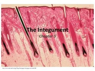
The Integument • the skin and all of its derivatives
Components • skin (epidermis, dermis, hypodermis) • derivatives (sweat glands, sebaceous glands, mammary glands, hair, nails, claws,hooves, horns, antlers, combs, wattles, and feathers)
Functions • protection - from drying out, from invasion by microorganisms, from UV light and from insults (mechanical, chemical or thermal) • sensation - for touch, pressure, pain and temperature • thermoregulation - decreases heat loss in cold temperatures; increases heat loss in hot temperatures • metabolic functions - energy stored in fat deposits; synthesis of vitamin D
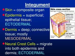
Structure of the Skin • Three distinct layers can be seen in the skin: • Epidermis - consists of keratinizing stratified squamous epithelium • Dermis - consists of fibroelastic connective tissue • Hypodermis - consists mostly of white adipose tissue (sometimes referred to as the subcutis)
Epidermis • Layers of the Epidermis • in order from outermost (surface) to innermost (deepest): • Stratum corneum - consists of the remains of keratinocytes; mostly composed of the protein, keratin • Stratum lucidum - present only in very thick skin; pale-staining layer of cells between the stratum corneum and stratum granulosum in which the dying keratinocytes contain a lot of keratin but are not completely replaced by it • Stratum granulosum - consists of keratinocytes containing large numbers of granules that contribute to the process of keratinization
Epidermis (Contd.) • • Stratum spinosum- consists of large, polyhedral keratinocytes that are actively • synthesizing keratin which is inserted as tonofibrils into the area of the plasma membrane • beneath desmosomes that connect adjacent cells together. These "connections" or • desmosomes between cells in this layer help hold them together and result in the "spiny" • appearance of the cells that gives this layer its name.
• Stratum basale - consists of keratinocytes undergoing mitosis to produce the constant • supply of keratinocytes needed for replacement of the dead and dying cells in the more • superficial layers of the epidermis
Dermis • Two zones of the dermis: • • papillary zone - consists of loose areolar connective tissue containing collagen and fine • elastic fibers; connects the epidermis to the thicker and denser reticular zone of the • dermis • • reticular zone - contains dense, irregular and coarse collagen fibers and thick elastic • fibers interspersed with fibroblasts and blood vessels and nerves
Glands in the skin. • Several different types of glands are located in the dermis of the skin serving a variety of • functions. • • Sebaceous glands • • Apocrine sweat glands • • Merocrine (= eccrine) sweat glands
Sebaceous Glands • The epithelium of this gland is an outgrowth of the external root sheath of • the hair follicle and the gland empties its oily product directly into the follicle itself. The glands • are of a branched acinar type and produce a lipid product called sebum that serves to reduce the • entry of microorganisms into the body through the skin, to lubricate the hair and preventing it • from drying out. The secretory cells die and become part of the product; a holocrine mode of • secretion. These glands are not found in hooves, foot pads, claws or horns.
Apocrine Sweat Glands • These glands are coiled, tubular glands with a large lumen and a duct • connecting it to an adjacent hair follicle. These glands secrete a viscid, milky product and are • analogous to odiferous glands of many mammals. Once thought to use the apocrine mode of • secretion, it is now known that their mode of secretion is more like that of the merocrine sweat • glands. These glands are the primary sweat gland of domestic animals and are especially • prominent in the horse.
Merocrine Sweat Glands • These glands are unbranched tubular in form and appear as a mass • of tubules in cross section. They are plentiful in the upper regions of the fatty hypodermis. They • secrete a watery product that is hypotonic to the plasma. It is the evaporation of this secretion on • the surface of the skin that aids in thermoregulation. These are sometimes called eccrine sweat glands.
Hair • General structure of hair and associated structures: • Hair shaft: the part of the hair above the surface of the skin • Hair root: the part of the hair below the surface • Bulb: an enlarged, hollow portion at the base of the root • Hair papilla: projection of dermis into center of the bulb • Follicle: the indentation in the skin within which the root lies
Layers of Hair • Cuticle: the outermost layer. Single layer of flattened, keratinized cells. Overlap like shingles, with free edge distally. • Cortex: the thickest, intermediate layer. Consists of several layers of keratinized cells containing hard keratin. • If hair is colored, these cells contain pigment. Cells held together by desmosomes. • Medulla: central core; loosely packed cuboidal cells. The structure and organization of the cuticle and medulla cells are species-specific.
Hair follicles • The hair follicle is the structure that anchors the hair in the dermis and produces the hair itself. It • is composed of five layers of epithelial cells arranged concentrically • The inner three layers form the hair shaft through a process of keratinization while the outer two layers form the hair sheath. • cells in the innermost layer form the medulla of the hair or core of the hair shaft • cells in the next layer form the cortex that makes up most of the hair • cells in the third layer form the cuticle on the surface of the hair
4. cells in the fourth layer make up the internal root sheath • 5. cells in the fifth or outermost layer form a layer called the external root sheath that does • not take part in hair formation • The external root sheath is separated from the surrounding connective tissue by a thick • basement membrane known as the glassy membrane.
Types of follicles • Hair follicles can be classified in two ways: based on their size, i.e., diameter and based on their organization. • Based on size (diameter): • Primary hair follicle: large having sweat gland, sebaceous gland and arrector pili muscle; ex. overcoat or guard hairs in dogs • Secondary hair follicle: smaller, lacking sweat glands and arrector pili muscle; ex. underhair
Based on Organization: • Simple follicle: a single hair from one follicle • Compound follicle: cluster of several follicles with several hairs emerging from one opening onto surface of skin
The Hoof • The equine foot includes the hoof, dermis, first, second and third phalanges and associated • structures. The hoof itself is the cornified layer of the epidermis, lacking the stratum granulosum • and stratum lucidum. It is important to understand the histology of the hoof because a disease • involving the epithelium of the foot, called laminitis, is the most devastating clinical disease of the foot.
The peculiar histology of the hoof is formed from special relationships between the dermis (or corium) and the overlying epidermis. • In some places, the dermal papillae and epidermal pegs are • confluent forming apparent layers, i.e., they are laminar or consist of lamellae; in other places they are more typical. • It is this lamellar interaction between the epidermis and dermis that gives the hoof its strength.
The wall of the hoof is that part of the hoof which is visible when the foot is on the ground, and • it can be divided into three layers. From outside to inside, they are the stratum externum • (tectorium), the stratum medium, and the stratum internum (lamellatum).
The Wall of the Hoof • The stratum externum or tectorium is an extension of the perioplic epidermis and is composed of cornified epithelial cells which appear as a soft, white, shiny material. This tissue attaches the hoof to the epidermis of the skin of the foot. • The stratum medium or coronary epidermis is composed of prominent tubular and intertubular horn, and this layer comprises the bulk of the wall of the hoof. • The stratum internum or stratum lamellatum is the epidermis in the laminar region.
Stratum Lamellum • This layer is made of nontubular horn which fuses with the stratum medium and helps hold the • wall to the foot. In this region, the dermal papillae and epidermal pegs form elongated ridges • oriented perpendicular to the ground. These ridges are formed from primary and secondary • laminae - the secondary laminae being oriented at close to a right angle to the primary laminae.
There are about 600 primary laminae and about 100-200 secondary laminae for each primary lamina. • This system of interdigitating primary and secondary laminae provides the tight bond between the wall of the hoof and the underlying dermis. • Thus, damage to the laminae leads to disruption of this interdigitating system which results in separation of the hoof wall from the dermis and phalanx beneath it.
Laminitis (acute laminar degeneration) is an inflammation of the laminae within the hoof. • Many pathophysiologic mechanisms are thought to cause laminitis, among them vasoconstriction within the digit, perivascular edema, arteriovenous shunting of blood at the level of the coronary band, venoconstriction and microthrombosis. • These lead to less than normal perfusion of blood to the digit resulting in ischemia, edema and eventually necrosis of the laminae.