Integument
Integument. Ms. Hughes Bio II. Integumentary System (Skin ). the skin has a surface area of 1.5 to 2 square meters, weighs 9 to 11 pounds (7% of total body weight in the average adult), and varies in thickness from 1.5 mm – 4 mm every square centimeter (cm) of the skin contains:


Integument
E N D
Presentation Transcript
Integument Ms. Hughes Bio II
Integumentary System (Skin) • the skin has a surface area of 1.5 to 2 square meters, weighs 9 to 11 pounds (7% of total body weight in the average adult), and varies in thickness from 1.5 mm – 4 mm • every square centimeter (cm) of the skin contains: • 70 cm of blood vessels, 55 cm of nerves, 100 sweat glands, 15 oil glands, 230 sensory receptors, and about 500,000 cells that are constantly dying/being replaced • pliable, tough, waterproof, insulates, cushions • without our skin, we would quickly fall prey to bacteria and perish from water and heat loss
protects the body from • mechanical damage (bumps and cuts) • chemical damage (acids, bases, poisons) • thermal damage (heat and cold) • UV radiation (sunlight) • invasion (bacterial)
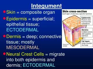
capillary network and sweat glands regulate heat loss • mini-excretory system (skin loses urea, salts, and water during sweating) • manufactures several proteins important in immunity • synthesizes vitamin D from sunlight and cholesterol • contains sensory receptors for touch, pressure, temperature, and pain • two distinct regions • epidermis and dermis • usually firmly connected but friction/burns may cause them to separate and form a blister
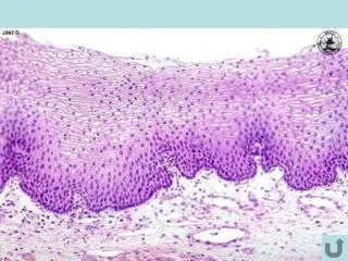
Epidermis • avascular • thick to prevent water loss • different types of cells
keratinocytes • produce keratin • protective properties • arise in the deepest part of the epidermis • undergo almost continuous mitosis • reach free surface of skin, they are dead, scalelike structures • (keratin-filled plasma membranes) • millions of these dead cells rub off every day • (we have a totally new epidermis every 35 to 45 days)
Melanocytes • specialized epithelial cells that synthesize the pigment melanin • touch all the keratinocytes • melanin granules accumulate on superficial, or "sunny," side of keratinocyte nucleus • forms a pigment shield to protect the nucleus from the damaging effects of UV radiation • same relative number of melanocytes • skin color due to differences in melanocyte activity • freckles/moles are where melanin is concentrated in one spot
Basal layer aka stratum germinativum (“growing layer”) • deepest • attached to the underlying dermis and receive nourishment • single row of cells representing the youngest keratinocytes • rapid division of these cells push daughter cells upward • alternate name, stratum basale (“bottom layer”) • 10% to 25% melanocytes
stratum spinosum (“prickly layer”) • weblike system of intermediate filaments • tension-resisting bundles of keratin filaments • keratinocytes in this layer are somewhat flattened and irregular in shape
stratum granulosum (“granular layer”) • three to five cell layers • nuclei/organelles begin to disintegrate which makes keratinocytes flat granules of keratin accumulate which helps in slowing water loss • superficial to this layer epidermal cells are too far from dermal capillaries • adequate nourishment is not received and they die
stratum lucidum (“clear layer”) • appears as a thin translucent band just above the stratum granulosum • clear, flattened, dead keratinocytes • present only in thick skin
stratum corneum (“horny layer”) • 20 to 30 cell layers thick • accounts for up to three-quarters of the epidermal thickness • keratin and thickened plasma membranes protect the skin against abrasion and penetration • glycolipids between cells waterproofs this layer • cornified/horny cells shed from the scalp and flakes slough off dry skin • average person sheds 40 pounds of these skin flakes in a lifetime
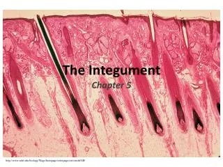
Dermis • vascularized, contains a rich nerve supply, is a shock absorber, and anchors the skin • nutrients reach the epidermis by diffusion • two major layers – papillary and reticular
Papillary Layer • fibers form a loosely woven mat heavily invested with blood vessels • superior surface has nipple-like projections called dermal papillae • contain capillary loops, nerve endings (pain receptors), and touch receptors • larger mounds called dermal ridges produce looped/whorled ridges on the epidermal surface • epidermal ridges increase friction and enhance the gripping ability of the fingers and feet • lots of sweat pores on ridges that leave unique patterns (fingerprints)
Reticular Layer • deeper layer containing sweat glands, oil glands, and blood vessels (80% of the dermis) • has deep pressure receptors and lots of phagocytes • has collagen fibers (toughness and attracts water to help the skin stay hydrated) and elastin fibers • (provides elasticity) - both are lost with age
Subcutaneous Tissue • deep to the dermis • known as the hypodermis or superficial fascia • anchors the skin to underlying organs • shock absorber and insulator for the deeper tissues
Skin Color • 3 pigments contribute to skin color: melanin, carotene, and hemoglobin
melanin • only melanin is made in the skin • ranges in color from yellow to reddish-brown to black • stimulated to greater activity when we expose our skin to sunlight • prolonged sun exposure causes a substantial melanin buildup
carotene • yellow to orange • accumulate in the stratum corneum and fatty tissue of the hypodermis • color is most obvious in the palms and soles, where the stratum corneum is thickest • hemoglobin • pinkish hue of fair skin reflects the red color of oxygenated hemoglobin
Appendages of the Skin • made in the epidermis - sweat glands, sebaceous (oil) glands, hair follicles/hair, and nails
sweat (sudoriferous) glands • formed by stratum germinativum and pushed deep into the dermis • entire skin surface except the nipples and parts of the external genitalia • 2.5 million per person
eccrine glands • more numerous • abundant on the palms, soles of the feet, and forehead • duct extends to open in a funnel-shaped pore • secretion is commonly called sweat • 99% water, with some salts, vitamin C, antibodies, traces of metabolic wastes, lactic acid, and small amounts of ingested drugs
pH between 4 and 6 • major role is to prevent overheating of the body • heat-induced sweating begins on the forehead and then spreads over the rest of the body • emotionally induced sweating (cold sweat brought on by fright, embarrassment, or nervousness) begins on the palms, soles, and armpits and then spreads to other body areas
apocrine glands • confined to the axillary and genital areas • larger than eccrine glands • ducts empty into hair follicles • same basic components as true sweat, plus some fatty substances and proteins • viscous with a milky or yellowish color • secretion is odorless • bacteria on skin use fat/proteins for nutrients and cause it to develop the musky body odor • begin to function at puberty • little role to play in thermoregulation
2 other specialized apocrine glands • ceruminous glands • found in the lining of the external ear canal • secrete a rather sticky substance called cerumen, or earwax • thought to deter insects and block entry of foreign material • mammary glands • specialized sweat glands • secrete milk
3. sebaceous (oil) glands • formed by stratum germinativum and pushed deep into the dermis • found all over the body except palms and soles • small on the body trunk and limbs, but large on the face, neck, and upper chest • oily secretion called sebum secreted into a hair follicle or to a pore on the skin surface • softens/lubricates hair and skin, prevents hair from becoming brittle, and slows water loss from skin • important bactericide
3. sebaceous (oil) glands • central cells of the gland accumulate lipids until they burst • stimulated by hormones - inactive during childhood • if blocked by accumulated sebum, a whitehead forms • a whitehead dries/darkens to form a blackhead • acne is an active inflammation of the sebaceous glands accompanied by pimples • caused by bacterial infection
4. hair follicles/hair • follicles extend from the epidermal surface into the dermis (hypodermis in the scalp) • deep end of the follicle is expanded, forming a hair bulb with sensory nerve endings (root hair plexus) • papilla (nipple-like bit of tissue) containing a knot of capillaries, protrudes into the hair bulb • wall thins as it approaches the hair bulb so that only a single layer of stratum germinativum cells covers the papilla to supply nutrients to the growing hair • the growth zone (matrix) in the hair bulb includes cells that actively divide to produce hair • (often triggered by chemical signals)
4. hair follicles/hair • associated with each hair follicle is a bundle of smooth muscle cells called an arrectorpili • contraction pulls the hair follicle into an upright position & dimples the skin surface (goose bumps) • bending the hair can also stimulate nerves (sensing an insect crawling over your skin) • millions of hairs are scattered over nearly all of the body • about 100,000 of them in the scalp (lose an average of 90 scalp hairs daily) • life span of hairs varies but follicles remain active for years (average is four) • eyebrow follicles remain active for 3-4 months • why eyebrows are never as long as the hairs on your head • rate of hair growth about 2 mm per week
4. hair follicles/hair • hair pigment is made by melanocytes at the base of the hair follicle • gray or white hair results from decreased melanin production • lips, nipples, parts of the external genitalia, and thick-skin areas (palms/soles) totally lack hair • hair on the scalp guards the head against physical trauma, heat loss, and sunlight • eyelashes shield the eyes • nose hairs filter large particles like lint and insects from inhaled air • hairs consist largely of flexible cells produced by hair follicles • as it grows, the older part of the hair is pushed upward, and its fused cells become increasingly keratinized and die
4. hair follicles/hair • hard keratin is tougher, more durable, and individual cells do not flake off • chief regions of a hair are the shaft (projects from skin), and the root (embedded in the skin) • cross sectional shape of the shaft determines straight or curly hair type: • flat/ribbonlike shaft - hair is kinky • oval shaft - hair is smooth and silky (maybe wavy) • round shaft - hair is straight and tends to be coarse
4. hair follicles/hair • classified as vellus or terminal • vellus • body hair of children and adult females • pale, fine • terminal • coarser, often longer hair of the eyebrows and scalp • body hair of adult males • appear in the axillary and pubic regions at puberty of both sexes • influenced by nutrition, hormones, and conditions that increase local dermal blood flow
4. hair follicles/hair • hair grows fastest from the teen years to the 40s - then growth slows (age-related atrophy) • leads to hair thinning and some degree of baldness • much less dramatic in women • coarse terminal hairs are replaced by vellus hairs • true (frank) baldness is male-pattern baldness and is genetically determined • delayed-action gene switches on in adulthood and changes the response to testosterone • follicular growth cycles become short (many hairs never emerge from follicles before shedding)
5. Nails • scalelike modification of the epidermis that contains hard keratin • forms a clear protective covering on the dorsal surface of the distal part of a finger or toe • each nail has a free edge, a body (visible attached portion), and a proximal root (embedded in the skin) • lateral/proximal borders are overlapped by skin folds called nail folds (proximal nail fold is the cuticle) • deeper layers of the epidermis (stratum germinativum) extend beneath the nail as the nail bed • thickened proximal portion of the nail bed, called the nail matrix, is responsible for nail growth • pink color due to underlying capillaries • some melanin can be seen thru the nail if the skin color is dark • white crescent over matrix is lunula
Homeostatic Imbalances of the Skin • the skin can have more than 1000 different disorders (allergies, bacterial, viral, fungal, burns, cancers) • athlete’s foot (tineapedis) • red, peeling skin between or underneath the toes (usually but not always itchy) • cold sores • herpes virus that localizes in a mucosal cutaneous nerve as a small blister that itches and stings • remains dormant until activated by emotional upset, fever, or UV radiation • contact dermatitis • itching, redness, and swelling of the skin due to chemical irritants • burns • tissue damage and cell death caused by heat, electricity, UV radiation, or chemicals • body loses fluids through seepage from wound leading to dehydration • dehydration leads to renal failure and circulatory shock • volume of fluid loss can be estimated by the “rule of nines”
the body is divided into 11 areas each representing 9% plus 1% for the perineum
burned skin is sterile for 24 hours before bacteria/fungi begin invading thru damaged areas • 1st degree burns – only the epidermis is damaged, redness/swelling occur, regeneration • 2nd degree burns – epidermis/upper dermis is damaged, blisters develop, regeneration • 3rd degree burns – destroys entire thickness of skin, burned areas appear white/black, no regeneration
Skin Cancer • single most common type of cancer • cause of most are unknown but the most important risk factor is UV exposure • most are benign and do not spread (warts) • some are malignant
basal cell carcinoma • least malignant, most common, slow growing • stratum germinativum does not form keratin and spread into the dermis • easily removed with high recovery rate • squamous cell carcinoma • appears red and scaly, rapid growth • starts in stratum spinosum on scalp, ears, dorsal hand • chance for complete recovery if caught early
malignant melanoma • cancer of the melanocytes • begins as spontaneous cancer in pigmented areas (often from pigmented moles) • metastasizes rapidly into surrounding lymph/blood vessels • often fatal