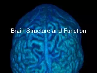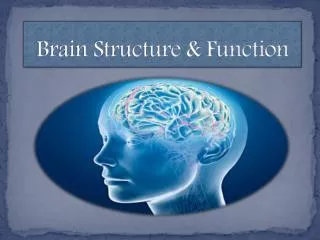
II. Brain Structure
720 likes | 896 Views
Brain, Mind, and Belief: The Quest for Truth. II. Brain Structure. If the brain were simple enough for us to understand, we would be too simple-minded to understand it. Anonymous. Brain Structure: Topics. Components of the Brain The cerebral cortex Neurons, axons, dendrites

II. Brain Structure
E N D
Presentation Transcript
Brain, Mind, and Belief: The Quest for Truth II. Brain Structure If the brain were simple enough for us to understand, we would be too simple-minded to understand it. Anonymous
Brain Structure: Topics • Components of the Brain • The cerebral cortex • Neurons, axons, dendrites • Synapses • Transmission of neural activity • Left brain and right brain
The nervous system • Central nervous system • Spinal cord • Brain • Peripheral nervous system • Motor and sensory neurons connected to the spinal cord
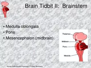
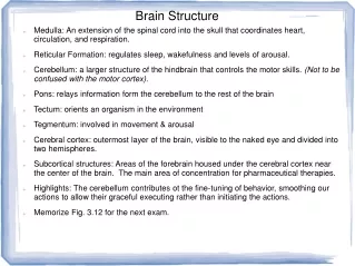
The brain • Medulla oblongata – Myelencephalon • Pons and Cerebellum – Metencephalon • Midbrain – Mesencephalon • Thalamus and hypothalamus – Diencephalon • Cerebral hemispheres – Telencephalon • Cerebral cortex • Basal ganglia • Basal forebrain nuclei • Amygdaloid nucleus • More..
The brain *Brain Stem • Medulla oblongata – Myelencephalon • Pons and Cerebellum – Metencephalon • Midbrain – Mesencephalon • Thalamus and hypothalamus – Diencephalon • Cerebral hemispheres – Telencephalon Alternative partition: Brain stem* Cerebellum Thalamus & hypothalamus Cerebral hemispheres
The brain • Medulla oblongata – Myelencephalon • Pons and Cerebellum – Metencephalon • Midbrain – Mesencephalon • Thalamus and hypothalamus – Diencephalon • Cerebral hemispheres – Telencephalon • Cerebral cortex • Basal ganglia • Basal forebrain nuclei • Amygdaloid nucleus
Thalamus and Cortex • The cortex is the area for • High-level information processing • Language • But the thalamus is also very important • Timing and coordination of cortical activity • Details not yet well understood • Metaphor: • The cortex is the orchestra • A very large orchestra • The thalamus is the conductor
Two hemispheres Right Left Interhemispheric fissure (a.k.a. longitudinal fissure)
Corpus Callosum Connects Hemispheres Corpus Callosum
Major Left Hemisphere landmarks Central Sulcus Sylvian fissure
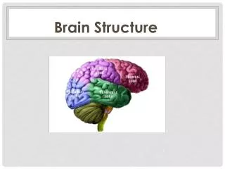
Major landmarks and the four lobes Central Sulcus Parietal Lobe Frontal Lobe Occipital Lobe Temporal Lobe Sylvian fissure
Primary motor and somatosensory areas Primary Somato- sensory Area Central Sulcus Primary Motor Area Sylvian fissure
Some terms.. • Fissures and sulci (the “grooves”) • Singular: sulcus – Plural: sulci • The major sulci are usually called fissures • Interhemispheric fissure • Sylvian fissure • Sometimes the term Rolandic fissure is used for the central sulcus • Gyri • Singular: gyrus – Plural: gyri
Alternatives terms for some fissures • Interhemispheric fissure • Also known as Longitudinal fissure • Sylvian fissure • Also known as Lateral sulcus • Central sulcus • Also known as Rolandic fissure
Primary Areas Primary Somato- sensory Area Primary Motor Area Primary Auditory Area Primary Visual Area
Divisions of Primary Motor and Somatic Areas Primary Somato- sensory Area Leg Primary Motor Area Trunk Arm Hand Fingers Mouth Primary Auditory Area Primary Visual Area
Higher level motor areas Primary Somato- sensory Area Actions per- Formed by leg Leg Actions performed by hand Trunk Arm Hand Actions performed by mouth Fingers Mouth Primary Auditory Area Primary Visual Area
Video of basic cortical anatomy http://www.youtube.com/watch?v=HVGlfcP3ATI&NR=1&feature=fvwp From Medical Legal Art (2009)
The brain operates by means of connections • Neurons do not store information • Rather they operate by emitting activation • To other neurons to which they connect • Via synapses • Proportionate to activation being received • From other neurons via synapses • Therefore, a neuron does what it does by virtue of its connections to other neurons • The first big secret to understanding how the brain operates
The cerebral cortex is a very large network • Made up of interconnected neurons • Very large • Dynamic • Changes take place in connection strengths • Every neuron is connected (directly or indirectly) to every other neuron • Therefore, all of the information in it has the form of a network • The information is in the connectivity • (stay tuned for further details)
Gray matter and white matter (coronal section) Gray matter White matter
Some brain quantities • The cortex accounts for 60-65% of the volume of the brain • But has only a minority of the total neurons of the brain • Surface of the cortex – about 2600 sq cm • That is, about 400 sq inches • Weight of cortex – • Range: 1,130 – 1,610 grams • Average: 1,370 grams • Brain mass nears adult size by age six yrs • Female brain grows faster than male during 1st 4 yrs • Thickness of cortex – (inf. from Mountcastle 1998) • Range: 1.4 – 4.0 mm • Average: 2.87 mm
Cortical Neurons • Cells, but quite different from other cells • Multiple fibers, branching in tree-like structures • Input fibers: Dendrites • Output fibers: Axons • Great variation in length of fibers • Short ones — less than one millimeter • Long ones — several centimeters • Only the pyramidal cells have such long ones
Cellular Communication:How to communicate with other cells • Method One (Nervous System): • Fibers projecting from cell body • Branching into multiple fibers • Input fibers – dendrites • Allow cell to receive from multiple sources • Output fiber – axon • Allows cell to send to multiple destinations • Method Two: • Circulation • Circulatory system • Endocrine system • Lymphatic system
Santiago Ramon y Cajal • 1852-1934 • Spanish neuroscientist • “The father of modern neuroscience” • Used microscope to examine brain tissue • Was skilled at drawing • Many of his drawings are still used today in teaching neuroscience • Nobel Prize in Medicine, 1906
Some quantities relating to neurons • Number of neurons • In cortex: ca. 27 billion (Mountcastle) • Beneath 1 sq mm of cortical surface: 113,000 • Synapses • 440 million synaptic terminals/mm3 in visual area • Each neuron receives avg 3,400 synaptic terminals
Formation of neurons in the fetus • 500,000 neurons are formed per minute in the developing fetus (from a program on PBS, 2002) • By 24 weeks, the brain has most of its neurons • Checking: • 500,000 per minute • 30 million per hour • 720 million per day • 5 billion per week • 96 billion in 24 weeks • Checks!
Brains of the young and very young • At about 7 months, a child can recognize most sound distinctions of the world’s languages • By 11 months the child recognizes only those of the language of its environment • At 20 months the left hemisphere is favored for most newly acquired linguistic information • Brain mass nears adult size by age six yrs • Female brain grows faster than male during 1st 4 yrs
Connecting fibers of pyramidal neurons Apical dendrite Basal dendrites Axon
Types of cortical neurons • Cells with excitatory output connections (spiny) • Pyramidal cells (about 70% of all cortical neurons) • Spiny stellate cells • Cells with inhibitory output connections (non-spiny) • Large basket cells (two subtypes) • Columnar basket cells • Double bouquet cells • Chandelier cells • Others
Pyramidal neurons Microelectronic probe About 70% of cortical neurons are of this type
Structure of pyramidal neuron Apical dendrite Cell body Axon Myelin
Neuronal Structure and Function:Connectivity • White matter: it’s all connections • Far more voluminous than gray matter • Cortico-cortical connections • The fibers are axons of pyramidal neurons • They are all excitatory • White since the fibers are coated with myelin • Myelin: glial cells • There are also grey matter connections • Unmyelinated • Local • Horizontal, through gray matter • Excitatory and inhibitory
Neuronal fibers • Estimated average 10 cm of fibers per neuron • A conservative estimate • Times 27 billion neurons in cortex • Amounts to 2.7 billion meters of neural fibers in cortex (27 billion times 10 cm) • Or 2.7 million kilometers – about 1.68 million miles • Enough to encircle the world 68 times • Seven times the distance from the Earth to the moon Big lesson: Connectivity rules!
Pyramidal neurons and their connections • Connecting fibers • Dendrites (input): length 2mm or less • Axons (output): length up to 10 cm • Synapses • Afferent synapses: up to 50,000 • From distant and nearby sources • Distant – to apical dendrite • Local – to basal dendrites or cell body • Efferent synapses: up to 50,000 • On distant and nearby destinations • Distant – main axon, through white matter • Local – collateral axons, through gray matter
Interconnections of pyramidal neurons Input from distant cells Input from neighboring columns Output to distant cells
Synapses • The connections between neurons • Neurotransmitters cross from pre-synaptic terminal to post-synaptic terminal • Synaptic cleft – about 20 nanometers
Quantity of synapses in the cortex • Synapses of a typical pyramidal neuron: • Incoming (afferent) – 50,000 (5 x 10⁴) • Outgoing (efferent) – 50,000 • Number of synapses in cortex: • 28 billion neurons (Mountcastle’s estimate) • i.e., 28 x 109 • Synapses in the cortex (do the math) • 5 x 104 x 28 x 109 = 140 x 1013 = 1.4 x 10¹⁵ • Approximately 1,400,000,000,000,000 • i.e., over 1 quadrillion
Release of neurotransmitter Presynaptic terminal releases neurotransmitter
Video of Synaptic Transmission http://www.youtube.com/watch?v=HXx9qlJetSU&feature=related By Jokerwe
Connections to other neurons • Excitatory • Pyramidal cells and spiny stellate cells • Output terminals are on dendrites or cell bodies of other neurons • Neurotransmitter: Glutamate • Inhibitory • All other cortical neurons • Output terminals are on cell bodies or axons of other neurons • Neurotransmitter: GABA • GABA: gamma-aminobutyric acid
Inhibitory connections Axosomatic Axoaxonal
Myelin (and other features) Dendrite Axon terminal Node of Ranvier Soma Schwann cell Myelin sheath Nucleus
Integration of neural inputs • Takes place at the axon hillock • Excitatory inputs are summed • Inhibitory inputs are subtracted • Result of this summation is the amount of incoming activation • Determines how much activation will be transmitted along the axon (and its branches), hence to other neurons • Degree of activation is implemented as frequency of spikes
Spread of activation • Activation moves across links • At the small scale • from neuron to neuron • At larger scale, across multiple links • In vision • From retina to conceptual area of cortex • In speech production, • from meanings to their expression • For a listener, • From expression to meaning