Download
1 / 30
310 likes | 457 Views
Explore the intricate details of the brain's gross anatomy, including dura mater, meninges, lobes, and compartments. Study how the cerebrum, cerebellum, and brainstem are organized within the cranium. Gain insights into the subarachnoid space, gyri, and sulci of the cerebrum. Delve deeper into the midbrain, pons, medulla, and cerebellum in the posterior cranial fossa. Enhance your knowledge of critical brain structures and their functions with this comprehensive guide.

E N D
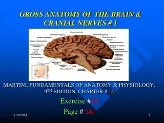
GROSS ANATOMY OF THE BRAIN Dr. G.R. Leichnetz
Craniectomy Exposes the Dura Mater The dura mater isthe outermost of the connective tissue coverings of the brain, the meninges. It is firmly attached to the inside of the skull, and its outer layer actually constitutes the periosteum of the inside of the skull. Thus, there is no epidural space in the cranium. The dura mater is supplied by branches of the middle meningeal artery.
The inner layer of the dura mater gives rise to partitions (dural reflections) that create two major compartments in the cranial cavity. The falx cerebri separates the two cerebral hemispheres. The tentorium cerebelli forms a roof over the posterior cranial fossa, separating its contents (cerebellum & brainstem) from the occipital lobes of the brain. Contains the cerebrum Falx cerebri Contains the cerebellum & brainstem Tentorium cerebelli Supratentorial Compartment- contains cerebrum Infratentorial Compartment (Posterior fossa)-contains cerebellum and brainstem Posterior fossa
When the dura mater is reflected it reveals the “leptomeninges” (arachnoid membrane + pia mater). The arachnoid is attached to the pia by arachnoid trabeculae. The space between the arachnoid and pia is the subarachnoid space. The subarachnoid space contains CSF which is reabsorbed into the systemic circulation thru arachnoid villi which project into the superior sagittal sinus. The cerebral arteries and veins run on the surface of the pia mater on the floor of the subarachnoid space. Arachnoid villi
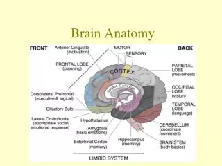
The surface of the cerebrum has gyri separated by sulci (grooves). The cerebrum has five lobes: frontal, parietal, temporal, occipital, and insular. The central sulcus separates the frontal and parietal lobes (note two vertical gyri, pre- and post-central). The lateral sulcus separates the frontal & parietal lobes from the temporal lobe. Parietal Lobe Frontal Lobe Occipital Lobe Temporal Lobe Insular Lobe
Cerebral Hemisphere Lateral Aspect Central sulcus Parietal Lobe Frontal Lobe Occipital Lobe Temporal Lobe Lateral sulcus Pre-occipital notch
Superior transverse temporal gyri (Primary Auditory Cortex) Insular Lobe Opening the lateral sulcus reveals the insular lobe. Insular Lobe
Lateral Aspect: Frontal Lobe Precentral gyrus Central sulcus Superior frontal gyrus Pars opercularis, inferior frontal gyrus Middle frontal gyrus Inferior frontal gyrus Pars triangularis, inferior frontal gyrus Pars orbitalis, inferior frontal gyrus
Lateral Aspect: Parietal Lobe Postcentral gyrus Central sulcus Superior parietal lobule Intraparietal sulcus Inferior parietal lobule Angular gyrus Supramarginal gyrus Lateral sulcus
Lateral Aspect: Temporal Lobe Superior temporal sulcus Lateral sulcus Superior temporal gyrus Middle temporal gyrus Inferior temporal gyrus
Ventral (Inferior) Aspect Frontal lobe Diencephalon(hypothalamus) Temporal lobe Midbrain Pons Medulla Cerebellum The brainstem (midbrain, pons, medulla) and cerebellum occupy the posterior cranial fossa.
Inferior Aspect, Frontal Lobe Olfactory bulb Orbitofrontal gyri Olfactory tract
Inferior Aspect: Temporal Lobe Middle temporal gyrus Parahippocampal gyrus Parahippocampal gyrus Uncus Midbrain Uncus Occipitotemporal (fusiform) gyrus Modified From: Haines Inferior temporal gyrus
Medial Aspect/ Cerebral Hemisphere (Mid-Sagittal Section) Central sulcus Parietooccipital sulcus Parietal Frontal Occipital Cerebellum Brainstem The central sulcus and parietooccipital sulcus are used to delineate lobes on the medial aspect of the hemisphere.
Medial Aspect: Frontal & Parietal Lobes Central sulcus Paracentral lobule Precuneus Superior frontal gyrus Cingulate gyrus Corpus callosum Parietooccipital sulcus
Medial Aspect: Occipital Lobe Parietooccipital sulcus Cingulate gyrus Precuneus Splenium, corpus callosum Cuneus gyrus Lingual gyrus Calcarine fissure Cerebellum
Midsagittal Aspect Paracentral lobule Parietooccipital sulcus Cingulate gyrus Cuneus gyrus Corpus callosum Thalamus Septum pellucidum Hypothalamus Midbrain Cerebellarvermis Calcarine sulcus IVth vent Pons Medulla
Mid-Sagittal: Closeup Pineal gland Septum pellucidum Fornix Thalamus Anterior commissure Superior colliculus Inferior colliculus Hypothalamus Midbrain Optic chiasm Mammillary body Pons Pituitary stalk (infundibulum)
Mid-Sagittal: Ventricular System Massa intermedia Lateral ventricle Fornix Pineal gland Choroid plexus thru interventricularforamen of Monro Cerebral aqueduct Anterior commissure Midbrain Fourth ventricle Lamina terminalis Lateral Recess & Foramen of Luschka Pons Third ventricle
Cerebellum: Anterior (Superior) Aspect Lateral hemisphere Posterior Lobe Vermis Primary Fissure Anterior Lobe Midbrain
Cerebellum, Posterior View Vermis Pyramis Posterior LobeHemisphere Uvula Tonsils of cerebellum Medulla Foramen of Magendie (opening in post. medullary velum from 4th ventricle)
Cerebellar Vermis Primary fissure Anterior lobe vernis Flocculus- hemispheric portion of the flocculonodular lobe of the cerebellum Fourth ventricle Posterior lobe vermis Nodule (vermal portion of the F-N lobe) Prenodular fissure Tonsil
Cerebellar Peduncles The cerebellar peduncles connect the cerebellum to the three divisions of the brainstem: SCP to midbrain MCP to pons ICP to medulla. They carry major tracts into and out of the cerebellum. Anterior lobe hemisphere removed to reveal peduncles Superior cerebellar peduncle Midbrain Middle cerebellarpeduncle Pons
Ventral (Inferior) Aspect of the Brain All cranial nerves exit from the ventral aspect of the brain, except the trochlear nerve. Telencephalon- I Diencephalon- II From the brainstem: Mesencephalon- III, IV Metencephalon- V Myelencephalon- VI, VII, VIII, IX, X, XI, XII
Brainstem: Ventral Aspect Cranial Nerves Optic (II) Oculomotor (III) Trigeminal (V) Facial (VII) and Vestibulocochlear (VIII) Glossopharyngeal (IX) & Vagus (X) Nerves
Ventral Aspect: Diencephalon & Midbrain Optic Nerve Optic Chiasm Oculomotor Nerve (CN III) Optic Tract Tuber cinereum and pituitary stalk Trochlear Nerve (CN IV) Cerebral peduncle Mammillary bodies
Ventral Aspect: Brainstem, Pons & Medulla Trigeminal Nerve (CN V) Middle cerebellar peduncle Pons Abducens Nerve (CN VI) Medulla Vestibulocochlear Nerve (CN VIII) Glossopharyngeal (IX) and Vagus (X) Nerves Facial Nerve (CN VII) Hypoglossal Nerves (CN XII) Spinal Accessory Nerve (CN XI) Olive Pyramid Pyramidal Decussation
Dorsal Aspect of the Brainstem (with cerebral cortex and cerebellum removed) Thalamus Thalamus Midbrain Midbrain Pons Rhomboid fossa Medulla Barr
Superior colliculus The roof of the midbrain is the tectum. It contains four elevations, the corpora quadrigemina. (superior & inferior colliculi) Inferior colliculus Anterior medullary velum Superior cerebellar peduncle The floor of the fourth ventricle is the rhomboid fossa. The “rhomboid” can be divided into two triangles. The rostral triangle is over the pons. The caudal triangle is over the medulla. Pons Middle cerebellar peduncle FC Medulla Obex
Dorsal Aspect of the Medulla Obex Gracile tubercle Cuneate tubercle Fasciculus graciclis Tuberculum cinereum Fasciculus cuneatus Dorsal intermediate sulcus