Download
1 / 35
400 likes | 620 Views
Explore complex neural pathways of somatosensory, visual, auditory, and vestibular systems involving spinal, thalamic, and cortical structures. Understand sensory information processing and potential lesions affecting perception.

E N D
SENSORY PATHWAYS REVIEW Dr. G.R. Leichnetz
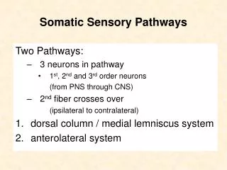
Postcentral gyrus/ paracentral lobule Somatosensory (GSA) Pathways- from the body: Lateral spinothalamic tract- pain & temperature Anterior spinothalamic tract- simple (crude) touch Dorsal column/ medial lemniscus- proprioception, vibratory sense, fine touch Ascend to terminate in the VPL nucleus of the thalamus, which then projects to the postcentral gyrus and paracentral lobule (leg). VPL Medial lemniscus Spinothalamic tracts Primary afferents
Tabes Dorsalis: Lesion of the Dorsal Columns) involving the fasciculus gracilis and/or cuneatus Loss of conscious proprioception, vibratory sense, fine touch, stereognosis below the level of the lesion. Positive Romberg sign. FG Lumbar spinal cord FG FC Cervical spinal cord
Syringomyelia Syrinx (cavitation) in cervical and upper thoracic spinal cord involves fibers crossing to join the anterior spinothalamic tract. Results in a bilateral segmental loss of pain and temperature at the level of the syrinx. Syringomyelia Lateral spinothalamic tract Syrinx LSTT is intact. Only crossing fibers at level of syrinx are affected. Thus segmental loss of pain & temp. Syrinx (cavitation) disrupts second-order spinothalamic axons, preventing them from joining the LSTT
Brown-Sequard Syndrome (hemisection of the spinal cord) involving the dorsal column (fasc. gracilis & cuneatus) and lateral spinothalamic tract. Ipsilateral loss of conscious proprioception, vibratory sense, and contralateral loss of pain and temp. sensation below the level of the lesion. FG CST LSTT FG= fasciculus gracilis CST= corticospinal tract LSTT= lateral spinothalamic tract
Brown-Sequard Syndrome (hemisection of the spinal cord) Dorsal columns: fasciculus gracilis & cuneatus Lateral spinothalamic tract LSTT FG Ipsilateral loss of conscious proprioception, vibratory sense (FG), and spastic paralysis (CST) Contralateral loss of pain & temperature (LSTT)
Somatosensory Pathways from head are related to the trigeminal nerve. Spinal Tract & Nucleus of V- pain & temperature Chief Sensory Nucleus of V- primarily touch (tactile) Mesencephalic Nucleus of V- proprioception from muscles of mastication. The spinal nucleus and chief sensory nucleus of V project thru contralateral (ventral) trigeminothalamic tract (trigeminal lemniscus) to terminate in the VPM nucleus of the thalamus, which projects to the head area of the postcentral gyrus. Postcentral gyrus, Head area VPM Trigeminal lemniscus Ophthalmic Trigeminal ganglion Maxillary Mandibular Spinal nucleus of V
Wallenberg’s (Lateral Medullary) Syndrome Ipsilateral loss of pain and temperature in head (spinal tract and nuc. of V) with contralateral loss of pain and temperature in the body (lateral spinothalamic tract). Loss of pain & temp. in ipsilateral face Loss of pain & temp. in contralateral body Occlusion of branches of Posterior Inferior Cerebellar Artery (PICA) to dorsolateral medulla Spinocerebellars (ataxia) Spinal tract & nucleus of V (loss of pain & temp./ head) Spinothalamic tracts (loss of pain & temp/ body)
Anterior Lobe, cerebellum Unconscious Proprioception Lower Limb-dorsal & ventral spinocerebellar tracts Upper limb- cuneocerebellar tract Upper limb Lower limb
Visual Pathway Retinal ganglion cells project their axons thru the optic nerve, chiasm and tract to the lateral geniculate nucleus. Ipsilateral projections to LGN layers 2,3,5; contralateral to 1,4,6. The LGN projects via the optic radiations to the cuneus and lingual gyri (area 17, primary visual cortex).
Visual Reflex (pupillary reflex) Pathway Pretectum Edinger- Westphal nucleus (OMC) Retinal ganglion cells project to the pretectum and superior colliculus. The pretectum projects to the Edinger-Westphal nucleus, which sends parasympathetic pregang. fibers to ciliary ganglion; postgang.’s to ciliary and sphincterpupillae muscles.. Ciliary ganglion Sphincter pupillae muscle
Visual Pathway Lesions: Optic Nerve Optic Chiasm Optic Tract Meyer’s Loop Cuneus Gyrus Lingual Gyrus
Receptor hair cells in the organ of Corti are on the peripheral processes of bipolar neurons of the spiral (cochlear) ganglion. The central processes travel with the auditory division of C.N. VIII to terminate in the dorsal and ventral cochlear nuclei.
Auditory System Second-order projections from the dorsal and ventral cochlear nuclei ascend crossed & uncrossed in the lateral lemniscus to the inferior colliculus and medial geniculate nucleus of the thalamus (with potential synapses in the superior olive, nuclei of the trapezoid body). The MGN projects to the superior transverse temporal gyri of Heschl, areas 41, 42, the primary auditory cortex. Primary auditory cortex MGN Inferior colliculus Lateral lemniscus
Primary vestibular fibers (from semicircular canals, saccule, utricle) in vestibular division of C.N. VIII terminate in all subdivisions of the vestibular complex. Vestibular complex Vestibular ganglion
Second-order vestibular fibers originating from the vestibular complex ascend in the medial longitudinal fasciculus (MLF) to terminate in the extraocular motor nuclei (III, IV, and VI). The basis of the vestibulo-ocular reflex To extraocular motor nuclei Medial longitudinal fasciculus (MLF) Vestibular complex Lesions of the vestibular division of VIII, vestibular complex, or MLF result in nystagmus.
Vestibulospinal Tracts Medial vestibulospinals (desc. MLF) only goes to cervical spinal cord (neck muscle motor neurons) The basis of the vestibulo-colic reflex. Lateral vestibulospinals extend the entire length of the spinal cord (affect posture/ equilibrium) Desc. MLF to cervical spinal cord LVST to entire spinal cord
Brainstem lesions involving vestibular structures (eg. vestibular division of C.N. VIII, vestibular complex, MLF, flocculonodular lobe of cerebellum) typically produce: nystagmus, rhythmic involuntary oscillation of the eyes; the eyes move slowly in one direction, and then jerk quickly back to the opposite side; or vertigo (dizziness, sense of room spinning); and difficulties with balance/equilibrium (postural problems).
Located in the pontocerebellar angle, a vestibular schwannoma can compress the vestibulocochlear nerve (C.N. VIII) with partial or complete deafness, tinnitis, vertigo, nystagmus, or facial nerve (C.N. VII) w weakness in ipsilateral face.
Olfactory Pathway (SVA): First-order bipolar neurons in the olfactory mucosa have central processes that synapse on mitral cells in the olfactory bulb. Mitral cells send their axons thru the olfactory tract to terminate in the primary olfactory cortex of the rostral temporal lobe (prepyriform & entorhinal cortex, and amygdala). Some olfactory projections go to the septum/basal forebrain region.
Taste Pathway (SVA): Facial N. (VII)- ant. 2/3 Glossopharyngeal N. (IX)- post. 1/3 Vagus N. (X)- epiglottis Cell bodies of taste neurons are in geniculate ganglion, and inf. ganglia of IX and X. SVA Petrosal ganglion (IX) GVA Nodosal ganglion (X) The solitary nucleus is the only visceral afferent nucleus in the brainstem) The rostral solitary nucleus is SVA (taste); caudal part is GVA (visceral sensation)
Taste Pathway (SVA) Solitary nucleus projects via tract which runs adjacent to the medial lemniscus to the VPMnucleus of the thalamus; with relay to the tongue region of the opercular part of the postcentral gyrus and insular cortex (consciousness of taste). Some ascending taste- related projections from the solitary nucleus terminate in the hypothalamus (effect on appetite). Taste area VPM Hypothalamus SVA Solitary nucleus
GVA fibers carrying visceral sensation (other than pain), have their cell bodies in the inferior ganglia of C.N. IX and X (petrosal & nodosal) and have their central connections with solitary tract and nucleus.Then the solitary nucleus projects to the hypothalamus and VPM. GVA visceral pain fibers travel with sympathetics, thru splanchnic nerves, thru sympathetic chain, white comm. rami, and dorsal root. Their cell bodies are in dorsal root ganglia. Central processes of unipolar neurons synapse in the dorsal horn of the spinal cord. VPM Hypothalamus Solitary Nucleus Thoracic splanchnics
Visceral pain fibers from thoracic & abdominal viscera travel in the reverse direction thru splanchnics (sympathetics) to the spinal cord. Their cell bodies are in the dorsal root ganglia. Their central processes synapse in the dorsal horn. While some visceral pain will travel with the lateral spinothalamic tract (neospinothalamic), most follows a multi-synaptic and slower ascending pathway through the brainstem to the thalamus (paleospinothalamic). Visceral Pain (GVA) DRG has GVA cell bodies Lateral spinothalamic tract Splanchnic nerves carry GVA fibers from gut GVA chemo- and mechano-receptors in the gut
Most GVA fibers carrying visceral sensation (other than visceral pain) travel with cranial nerves IX and X. GVA fibers from carotid sinus (blood pressure) and carotid body (blood gases), have their cell bodies in the inferior ganglion of C.N. IX (petrosal). GVA fibers from the gut have their cell bodies in the inferior ganglion of C.N. X (nodosal). Central connections with solitary tract and nucleus. General Visceral Sensation (GVA) CN IX Solitary nucleus Petrosal ganglion CN X The solitary nucleus is the only visceral afferent nucleus in the brainstem (GVA, SVA). Nodosal ganglion Carotid sinus Thoracic splanchnics Visceral pain
Ventral Aspect of the Brain All cranial nerves exit from the ventral aspect of the brain, except the trochlear nerve. Telencephalon- I Diencephalon- II From the brainstem: Mesencephalon- III, IV Metencephalon- V Myelencephalon- VI, VII, VIII, IX, X, XI, XII
Functional Components of Cranial Nerves GSA= receptors in skin & muscle GVA= receptors in gut & large blood vessels SSA= receptors of special senses SVA= receptors for taste & smell GSE= to muscles derived from somites (eg. extraocular and tongue muscles) SVE= to muscles derived from visceral arches (branchiomeric muscle) GVE= to smooth (gut, glands) & cardiac muscle (autonomic) I Olfactory- SVA (smell) II Optic- SSA (vision) III Oculomotor- GSE (extraocular), GVE (ciliary, sphincter pup.) IV Trochlear- GSE (extraocular, sup. oblique) V Trigeminal- GSA (pain, temp., head) , SVE (masticatory) VI Abducens- GSE (extraocular, lat. rectus) VII Facial- SVA (taste, ant. 2/3), SVE (facial), GVE (submaxillary & sublingual salivary glands) VIII Vestibulocochlear- SSA (audition) IX Glossopharyngeal- GVA (carotid sinus & body), SVA (taste, post. 1/3), SVE (stylopharyngeus), GVE (parotid saliv.gland) X Vagus- GVA (gut), SVA (taste, epiglottis), GVE (parasymp. to gut); SVE (laryngeal muscles) XI Spinal Accessory- GSE (sternocleidomastoid & trapezius) XII Hypoglossal- GSE(tongue)