Download
1 / 54
600 likes | 866 Views
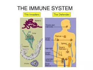
Introduction to the Immune System. Hilary Joyce Principal Clinical Scientist Immunology Birmingham Heartlands Hospital B9 5SS . Physical Barriers to Infection. Skin - a tough layer of keratinised cells defends the outside of the body

E N D
Introduction to the Immune System Hilary Joyce Principal Clinical Scientist Immunology Birmingham Heartlands Hospital B9 5SS
Physical Barriers to Infection • Skin - a tough layer of keratinised cells defends the outside of the body • The inside of the body is protected by mucosal membranes that secrete protective substances • e.g the lysosyme in tears and saliva is an anti-bacterial substance • e.g the acid pH of the stomach & vagina
The Immune System • The Innate or Non-Specific Immune Response • The Acquired, Adaptive or Specific Immune Response
Innate phylogenetically older quicker to act sends cytokine signals to the adaptive IR Adaptive did not evolve until the vertebrates specific for a particular pathogen memory cells are long lived The Immune Response
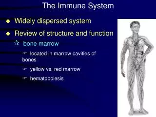
Cells of the Immune System • All the cells of the Immune System are leucocytes and originate in the bone marrow from a common precursor, the pluripotent stem cell. • Some cells are mature when they leave the marrow. • Other cells complete their differentiation outside the marrow.
Stem Cell Lymphoid cell line Myeloid cell line Polymorpho -nuclear leucocytes Monocytes B T NK Macrophages Neutrophils Eosinophils Basophils Specific Innate
Cells of the Innate Immune System • The main cells of the Innate Immune System are phagocytes - Macrophages and Neutrophils • These cells recognise structures which frequently occur on bacterial cells. • There is no development of long term immunity to a particular pathogen • NK cells are also part of the Innate IR
Phagocytes (1) • Play a major role in killing pathogens • Receptors on their cell surface that recognise molecules that are common to many bacteria. • Once bound to the receptor the pathogen can be phagocytosed and killed. • The cells can release cytokines to send messages to recruit other cells of the IS
Macrophages derived from circulating monocytes exist in the tissues often the first cells to detect and attack pathogens. long lived cells co-operate with lymphocytes Neutrophils most numerous white cell in the blood. remain in the circulation until they receive signals indicating invading microbes. Short lived cells Phagocytes (2)
Phagocytes (3) • Both macrophages and neutrophils phagocytose the bacteria and then kill them with destructive enzymes and free oxygen radicals. • Some bacteria e.g Listeria, M Tb have adapted to life in the macrophage • these then need activating to boost their ability to kill the bacteria.
Inflammatory Reaction • Inflammation is initiated by chemicals released by the phagocytes once they have encountered a pathogen. • Dilation of capillary walls allows • an increased blood supply (redness & warmth) • more leakage of plasma proteins (swelling & pain) • more neutrophils to leave the circulation and join in the fight
Cells of the Specific Immune System (1) • Lymphocytes (small) • They carry a specific antigen recognition molecule on their surface. • Recognition of their specific antigen drives differentiation and proliferation to produce a highly specific and effective clone.
B cells mature in the marrow carry Ig on the cell surface that act as receptors for their specific antigen Major role is to differentiate into plasma cells that produce antibodies T cells mature in the thymus antigen recognition structure is the T Cell Receptor (TCR) can mature into different types of effector cells Helper (Th) Cytotoxic (Tc) Cells of the Specific Immune System (2)
Primary and Secondary Lymphoid Tissue • Primary Lymphoid Tissue • sites where lymphocytes complete their maturation • Bone Marrow & Thymus • Secondary Lymphoid Tissue • Sites where the mature cells are stimulated to respond to their antigens • this is distributed throughout the body lymph nodes, spleen,Peyer’s patches, tonsils, appendix.
B cell Development • Very wasteful as the majority are eliminated • only 1 in 3 will produce functioning Ig after gene rearrangement • others may react with self antigen in which case they are either • removed by apoptosis • rendered anergic • Naïve B cells have a half -life of 3 -8 weeks
T cell Development • Aim to develop as wide a repertoire of TCR’s as possible • Immature T cells leave the marrow and travel to the thymus • Positive selection eliminates those cells whose TCR’s cannot react with self MHC • Negative selection removes those cells that would react too strongly with MHC • i.e. those that are potentially auto-reactive.
Principles of Adaptive Immunity • The Adaptive Immune System can produce millions of TCR’s & Ig’s that recognise different molecules • on infection only those lymphocytes that can recognise their antigen will respond.
B cells • B cells display ‘their’ Ig as the antigen receptor (sIg) • When stimulated they differentiate into effector cells i.e plasma cells • Plasma cells produce circulating Ig that is identical to the sIg (apart from the carboxy terminal end that anchors sIg into the B cell membrane.) • Ig’s can recognise a wide range of types of molecules
T cells • T cells display TCR as their antigen recognition protein. • When stimulated they become Cytotoxic or Helper T cells. • They secrete cytokines that recruit other cells of the IS • TCR’s only recognise short peptides.
Generation of Diversity (1) • The antibody repertoire of an individual could be as large as 1016 • A large majority of the human genome would be taken up coding for these molecules alone. • Immunoglobulins break the ‘One Gene = One Protein’ rule
Generation of Diversity (2) • The germ-line cells contain gene segments capable of encoding the antibody repertoire. • This diversity is created by : • Gene re-arrangements • Junctional diversity • Combinatorial diversity ( H & L chains) • Somatic hypermutation ---> affinity maturation
Generation of Diversity (3) The germ line cells contain the cells for all the possible genes
Generation of Diversity (4) During the development of B cells the V, D & J segments are cut and spliced by DNA recombination. During this somatic recombination a single gene of each type is brought together
Generation of Diversity (5) The light chains also contribute to the possible variations
Generation of Diversity (6) • The specificity of the TCR’s is determined in a similar manner. • This ability to cut and splice DNA is unique to lymphocytes. • These recombinations also introduce additional nucleotides not coded for in the germ line genes. • this Junctional diversity adds a factor of 3 x 10⁷ to the overall diversity
Antigen Recognition • B cells can recognise antigens via their sIg alone • T cells can only recognise antigen in association with a Major Histocompatibility Complex (MHC) molecule.
MHC Class 1 detected on all nucleated cells very highly polymorphic Tight fit for peptides of only about 9 aa consists of an a-chain of 3 domains associated with b-2 microglobulin MHC Class II seen only on the ‘professional antigen processing cells’ e.g macrophage slightly less polymorphic accepts peptides of up to 15 aa acids polymorphic b chain + a (monomorphic for DR) MHC
MHC & T cells • T cells have a requirement to recognise both the ANTIGEN and the MHC molecule. This is because the molecular structure of the MHC-Antigen complex is arranged so that some of the polymorphic amino acids of the MHC molecule are in direct contact with the TCR • Therefore T cell recognition of antigen is said to be MHC ‘restricted’.
MHC Class I present endogenously derived peptides. these can be either self or derived from viruses because MHC Class I is present on all cells any cell can interact with T cells if infected by a virus MHC Class II present exogenous antigen which has been phagocytosed and processed.eg. Bacteria This is performed by professional antigen presenting cells eg macrophages MHC & Antigens
Effector T cells • These are distinguished by cell surface markers • CD8 bearing cells are the Cytotoxic T cells & recognise MHC Class I.They kill cells infected with viruses • CD4 bearing cells are the Helper cells & recognise MHC Class II & secrete cytokines that activate other cells of the IS.
Th1 secrete cytokines that mainly activate the macrophages to phagocytose and kill extracellular pathogens Th2 secrete cells that help the effector B cells (plasma cells) to produce more antibodies Helper T cells Th1 & Th2 cells co-operate to eliminate pathogens
Immunological Memory • The Adaptive IS generally gives rise to immunity. • T & B cells differentiate after meeting ‘their’ antigen, although most cells produced are effector cells some ‘Memory’ cells that are long lived are also made. • On later encounters with the same pathogen the memory cells are activated
Complement (1) • Major defence mechanism against extracellular pathogens. • Series of proteins that aid the elimination of pathogens by • opsonisation • Cell lysis • Some parts can also recruit cells of the IS to help
Classical Pathway • The first component of the Classical Pathway of Complement is C1q. • This binds between adjacent Fc regions of Igs • C1r and C1s are added to form an active protease. • Then C4 is cleaved by the complete C1 complex. • C4a is released into the circulation • C4b binds to the cell surface • C1 can also cleave C2 in a similar manner • C4a is released into the circulation • C2b and C4b combine to form a C3 convertase
Lectin Pathway • Mannan Binding Protein is structurally very similar to C1q but it can combine directly to any mannose groups on the bacterial cell wall without the need for specific antibodies. • MASP is a protease capable of reacting with C4 & C2 resulting in the formation of C4bC2b, the same C3 convertase that is formed by the action of the Classical Pathway.
Alternative Pathway • C3 is an unstable molecule continually undergoing hydrolysis in the blood and lymph. This rate of hydrolysis can be accelerated in the presence of some pathogens. C3b then binds to Factor B making it susceptible to cleavage by Factor D. This creates the C3 convertase of the Alternative Pathway, C3bBb.
C3 • Central molecule in the Complement System. • Highly labile • Present in large amounts in serum (0.7-1.5g/l) • After action by the C3 convertases C3b is covalently bound to the surface of the target cell.
Assembly of the Membrane Attack Sequence • C3 convertases also bind to C3b to produce C5 convertases. • C5b initiates the formation of the Membrane Attack Complex • C6 & C7 bind to C5b, exposing a site on C7 that allows insertion into the lipid bi-layer • C8 binds and also inserts into the bi-layer and initiates polymerisation of C9
Small Complement Fragments • The fragments C3a, C4a & C5a can • act as chemotaxins to attract inflammatory cells such as neutrophils to the site of Complement Activation i.e. of infection • act as anaphylatoxins and induce degranulation of mast cells that releases histamine and other vasoactive substances that increase vascular permeability
Chemical Messengers synthesised by or Affecting Cells of the Immune System • Cytokines • proteins made by cells that that affect the behaviour of other cells • Lymphokines • cytokines made by lymphocytes also known as • Interleukins • numbered IL-1 to IL-20(ish & still counting) • Chemokines - small cytokines • Interferons • cytokines that help cells to resist viral infections • TNF - another family of cytokines • Cytotoxins - only produced by Cytotoxic T cells
Cytokines (1) • The same cytokine may be produced by more than one type of cell • The same cytokine may have more than one function • They may act on the cell that produced them - autocrine or on another type of cell- paracrine • They usually act locally and over a short period.
Cytokines (2) • Cytokines interact with a membrane bound receptor with a cytoplasmic tail • This interaction triggers a series of reactions that generally results in a change of gene expression within that cell
Immunoglobulins (1) • All antibodies are immunoglobulins although not all Igs can function as antibodies • The function of an antibody is to bind its antigen as tightly as possible and then direct it towards other components of the IS so that it can be destroyed.
Immunoglobulins (2) • Antibodies can be made against proteins, carbohydrates, lipids and nucleic acids. • but • Antibodies to nucleic acids and lipids can be found in autoimmune diseases. • Antibodies to small organic molecules can cause allergies to drugs
Immunoglobulins (3) Amino terminal Carboxyl terminal
Immunoglobulins (4) • Antibodies are glycoproteins made up of 2 heavy and 2 light chains. • Each pair of chains is identical. • They form the basic Y shape with a pair of antigen binding sites and a constant region. • The chains are held together by di-sulphide bridges. • The light chains are either k or l • In humans the ratio of k to l is 2:1
Immunoglobulins (5) • The Heavy chains are a, g, m, d or e • The sites and placing of the di-sulphide bridges and of the sugar groups varies with the class of heavy chain. • The biological functions are dependant on the class of Heavy chain e.g ability to cross the placenta or to fix complement. • IgM is pentameric • IgA is present in serum as a monomer and in secretions as a dimer.