Download

1 / 17
170 likes | 202 Views
Gel Electrophoresis. Exercise 5. Announcements. Post Lab 5 is due before your lab meets next week. Pre Lab 8 will be made available the week of March 16 and due before your Lab 8. LNA DNA/DNA Electrophoresis is assigned today and due next week before Exercise 6 (Exam 1 Review).

E N D
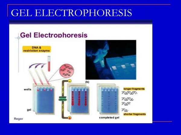
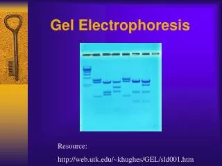
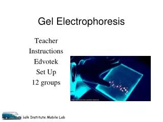
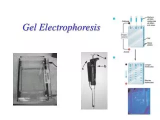
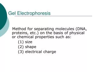
Gel Electrophoresis Exercise 5
Announcements • Post Lab 5 is due before your lab meets next week. • Pre Lab 8 will be made available the week of March 16 and due before your Lab 8. • LNA DNA/DNA Electrophoresis is assigned today and due next week before Exercise 6 (Exam 1 Review). • Sign up for Exam I. Exam I will take place during your lab period the Week of March 6. • There will be a Review Sheet for Exam I posted under Week 7. • Exam I covers material from Exercises 1-5. • The last day to drop MCB 151 is March 10.
Week 5 Itinerary • Find your samples from week 4 and store them on ice • Wear gloves • Check out kits • Pick up gel • Load and run gel • TA teaches • Take photos of gel (make 3 copies so you each have one for your notebook assignment) • Make sure you understand your photo before leaving
CAUTION • UV Light • UV light will damage your eyes
DNA Gel Electrophoresis • Negatively charged DNA is pulled toward the anode • Large pieces/fragments of DNA do not move as fast as smaller ones
- C B BH H +
- C B BH H +
- C B BH H +
- C B BH H +
- C B BH H +
- C B BH H We can convert migration distances into fragment size (kB) by using a standard curve (Size versus migration distance) +
- λ C B BH H Measure Known 6.5 kB 5.0 kB 4.0 kB 3.5 kB 3.0 kB 2.0 kB 1.0 kB 0.5 kB +
- λ C B BH H Known 6.5 kB 5.0 kB 4.0 kB 3.5 kB 3.0 kB 2.0 kB 1.0 kB 0.5 kB +
4 mm 10.0 6 mm 8 mm 5.0 9 mm 10 mm 3.0 2.0 13 mm 1.0 0.5 19 mm 0.3 0.2 24 mm 0.1 4 6 8 10 12 14 16 18 20 22 24 Standard Curve Size (kBases) Known Measured 6.5 kB 5.0 kB 4.0 kB 3.5 kB 3.0 kB Size (Kb) 2.0 kB 1.0 kB 0.5 kB Distance Migrated (mm)
4 mm 10.0 6 mm B B BH BH H H 8 mm 5.0 mm mm mm mm mm mm 9 mm 10 mm 3.0 2.0 13 mm 1.0 0.5 19 mm 0.3 0.2 24 mm 0.1 4 6 8 10 12 14 16 18 20 22 24 9 9 8 24 Size (kBases) Known Measured 6.5 kB 5.0 kB 4.0 kB 3.5 kB 3.0 kB Size (Kb) 2.0 kB 1.0 kB 0.5 kB Distance Migrated (mm)
Size of this fragment? H You need to make a “map” like this for your plasmid. B • What gel do you expect? (B, H, BH) • Be sure to include plasmid size on your map. Size of this fragment? Plasmid Size = ? kBases
Trouble Shooting (when gel bands are less than ideal) • Consider: • Different amounts of supercoiling • Partial Digests • Small fragments are harder to see (less DNA so less GreenGlo intercalating)