Download
1 / 79
790 likes | 931 Views
Explore the intricate functions of the cardiovascular system, including the role of blood vessels in maintaining homeostasis and transporting essential substances. Learn about the specialized endothelium and various types of blood vessels.

E N D
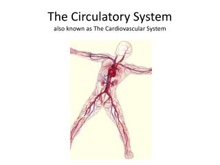
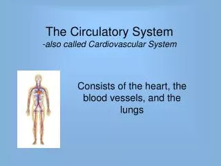
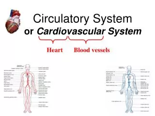
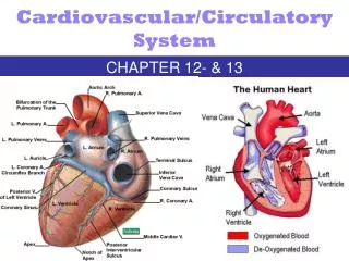
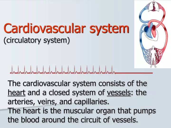
Cardiovascular system(circulatory system) The cardiovascular system consists of the heart and a closed system of vessels: the arteries, veins, and capillaries. The heart is the muscular organ that pumps the blood around the circuit of vessels.
Characteristics of c-v system • is an unique and complex hydraulic system • is a closed circle ("circulatory system") • is elastic
Function of c-v system • to maintain homeostasis and an optimal cellular environment. • transport function of oxygen, nutrients and waste products • lining endothelium is important for these functions; is waterproof and incoagulable
Endothelium is a specialized form of mesenchyme-derived epithelium simple squamous epithelium – one layer of flattened cells forms a thin, waterproof and antithrombogenic lining of all bloodvessels,heart and lymphatic vessels
Endothelial cells (1,2) in ELM ERY lumen Pinocytic vesicles Zonula occludens 2 1 blood Pinocytic vesicles tissue
Function of endothelium • the control of blood pressure by vasoconstriction and vasodilation, • blood clotting, • formation of new blood vessels (angiogenesis), • control of the passage of materials and the transit of white blood cells into and out of the blood, • in some organs, there are highly differentiated endothelial cells to perform specialized 'filtering' functions (renal glomerulus in kidney, blood-brain barrier, placental barrier).
Blood vessels are categorized by function • Arteries conduct blood away from the heart and have proportionately more smooth muscle and elastic tissue than veins of comparable size. • Arteries are commonly sub-categorized into elastic arteries (the largest one), muscular arteries (middle-sized), and arterioles. • Veins return blood to the heart. The composition of the wall varies among arteries and veins.
Bloodstream organization veins arteries venules arterioles postcapillaries precapillaries capillaries
Blood capillaries Network of the smallest, thin- walled vessels, situated between arterial and venous portion of circuit
Blood capillaries • diameter from about 8 µm (to 30-40 µm) • lumen is lined by 1-2 endothelial cell • reticular fibers surround the capillaries • capillary bed between arteries and veins • pericytes 3 types of capillariescontinuous fenestrated sinusoids
Function of capillaries (1) • respiratory gasses, nutrients and waste products change between blood and tissues The illustration shows satistied cells in well vascularized tissue
Function of capillaries (2) • allow the blood cells to pass throughout their wall into the connective tissue (by diapedesis) Neutrophils microphages Eosinophils Basophils heparinocytes Lymphocytes Monocytes macrophages
The smallest: cca 8 m The wall: - endothelium – 1-2 cells (zonulae occludentes and nexuses) - lamina basalis - pericytes - reticular fibers only allow small molecules, water and ions to diffuse Continuous capillaries Example of occurrence: muscle tisue, brain
Endothelial cells with fenestra („windows“) 70 nm , diaphragm (thinner than plasma membrane) boards fenestrum continuous basal lamina in the organs with quic and intensive metabolism and substances change allow small molecules and limited amounts of protein to diffuse Fenestrated capillaries Exampl of occurrence: intestinal villi, endocrine glands
Capillaries with pores • special type of fenestrated capillaries • not fenestra with diaphragm, but opened pores are in endothelium • in glomeruli of renal corpuscles Pores capillary lumen basal lamina
from 8 to 40 m endothelium – fenestra, pores and intercellular clefts; some cells are able to phagocyte incomplete basal lamina reticular fibers allow erytrhocytes and serum proteins to enter. Sinusoidal capillaries (sinusoids) Example of occurrence: liver, spleen, bone marrow
Remember! • Sinusoid = type of blood capillary (between arterial and venous part of bloodstream) • Sinus = venous sinus belong to venous, postcapillary part of bloodstream sinusoid vs. sinus
cytoplasmic processes around capillary, contain actin, myosin, tropomyosin their own basal lamina fuses together with that one of capillary Pericytes
12 – 40 m endothelium + LB, elastic + collagen fibers, smooth muscle cells precapillary sphincters to 200 m endothelium + LB, smooth muscle cells Precapillaries - Postcapillaries
Structure of blood vessel wall – generally – • tunica interna (intima) endothelium + subendothelial connective tissue ________membrana elastica interna__________ • tunica media smooth muscle tissue – circularly oriented ________membrana elastica externa__________ • tunica externa (adventitia) loose connective tissue + nerves + vasa vasorum (+ longitudinal smooth muscle – only in veins)
Endothelium Tunica interna <lungitudinally> Membrana elastica interna Tunica media <circularlly> (Membrana elastica externa) Tunica externa <longitudinally>
Tunica interna (intima) TI • endothelium • subendothelial connective tissue – thin layer of elastic + collagen fibers (longitudinally oriented)
Tunica media TM • consists of smooth muscle cells and elastic membranes in varying proportions (circularly oriented) • is thicker in arteries than in veins Compare aw – vw:
Tunica externa (adventitia) TA • fibrous connective tissue + smooth muscle cells in veins (logitudinally) • is thicker in vein; is the thickest layer in large veins [1] and veins of low limbs [2] • contains vessels and nerves (vasa et nervi vasorum) in large vessels 2 1
Structural differences between arteries and veins – generally: + + ++ (+) YES Membrana elastica int. (YES) thick thin (YES) Membrana elastica ext. NO thick thin
Compare the wall structure of artery and vein adventitia media (+ intima)
Arterial part of bloodstream According to diameter, morphological differences and ratio of elastic fibers and smooth muscle cells: • Arterioles < 0.5mm • Muscular arteries (small and middle-sized) 0.5 – 1 mm • Elastic arteries (large: aorta and arteries growing from aorta)
Arteriole • < 0.5mm The wall • TI: endothelium + subendothelium • membrana elastica int. • TM: smooth muscle cells (cca circular 5 layers) • TA: fibrocytes, reticular (+collagen) fibers
Muscular artery • TI: endothelium + subendothelium (with smooth muscle cells (longit.) • membrana elastica int. • TM: up to 40 layers of smooth muscle cells, elastic and collagen fibers • membrana elastica ext. • TA: loose connective tissue „circular“ „longitudinal“ TM+TA arrangement is spiral, but …
Elastic artery • TI: endothelium + subendothelium (100 m wide layer of connective t.) • TM: up to 40-60 layers of fenestrated elastic membranes, small amount of smooth muscle cells and reticular fibers • TA: loose connective tissue (+ vasa et nervi vasorum)
Different types of arteries • Arteriovenous anastomosis(artery contains smooth muscle cells in the wall before vein) • Arteries with intimal pillows(smooth muscle cells form pillows in t.media) – lumen can be closed by their contraction
Portal circulation:arterial or venous • two capillary systems side-by-side 2 3 1 capillaries vessel capillaries WHERE? 1: glomerulus eferent arteriole renal tubules capillaries in KIDNEY 2: GIT organs vena portae hepatic sinusoids in LIVER 3: hypothalamus hypophyseal vein adenohypophysis in HYPOPHYSIS
Venous part of bloodstream • Venules 0.2 – 1 mm • Small and medium sized veins 1 – 9 mm • Large veins(v. cava inf. et. sup. - the largest vein) • Valves - pocket-like duplication of endothelium scaffolded by elastic c.t. - protection against venous reccurence
< 0.2 - 1mm The wall TI: endothelium only TM: smooth muscle cells (cca circular 1-3 layers) TA: thick layer of loose connective tissue Venule
1 – 9 mm TI: endothelium + irregular layer of subendothelium + valves TM: irregular, thin layer of smooth muscle cells, elastic and collagen fibers TA: thick layer of loose connective tissue with smooth muscle cells Small and medium-sized venules Vein + artery Vein from lower part of body
TI: endothelium + subendothelium (+smooth muscle cells) TM: thin layer of connective tissue + reduced amount of smooth muscle cells TA: longitudinal bundles of smooth muscle cells in loose connective tissue (vasa et nervi vasorum) Large veins
The heart is the hardest working muscle in the human body. • Hollow muscular organ – blood pump • Rythmic contraction • Involuntary muscle
Pericardial sac:pericardium + epicardium PERICARDIUM Outer (parietal) layer Pericardial cavity EPICARDIUM Inner (visceral) layer Pericardial cavity - contains 15 – 50 ml of serous fluid serves as lubricans; - is lined with mesothelium
The wall of heart • Epicardium • Myocardium • Endocardium -------------------
Endocardium(homologous to intima of blood vessels) Consists of: • Endothelium • Subendothelium– thin connectivetissue layer • Elastic-muscular layer– dense c.t. (elastic fibers, smooth m. cells) • Subendocardium– c.t. + vessels, nerves and distal part ofconducting system (ventricular bundles and Purkinje fibers) Attention! Purkinje fibersPurkinje cells
Myocardium • cardiomyocytes „working“ „conducting“ • cells in right ventricle – natriuretic factor (when intravascular volume increases, this factor is released and causes natriuresis and diuresis in kidney) • atrial myocardium is thinner than ventricular • „left heart“ myocardium is thinner than „right heart“ • cords of cardiomyocytes are ended on heart skelston • damage of myocardium - infarction • low regeneration of myocardium – by scar (decreases function of heart muscle)
Heart skeleton Trigonum fibrosum sin. Trigonum fibrosum dx. Anulus fibrosus sin. Anulus fibrosus dx. Pars membranacea interventricularis