Download
1 / 19
190 likes | 325 Views
Hospital Virgen de los Lirios de Alcoy María Planelles. JoseLuis Losa. Jaime Vierna Valencia, 20 noviembre 2009. Historia Clínica. Hombre 67 años acude a urgencias desde Centro Salud por ictericia mucosa y cutánea No RAM Diabetes tipo II. Insuficiencia Renal Crónica

E N D
Hospital Virgen de los Lirios de AlcoyMaría Planelles. JoseLuis Losa. Jaime ViernaValencia, 20 noviembre 2009
Historia Clínica • Hombre 67 años acude a urgencias desde Centro Salud por ictericia mucosa y cutánea • No RAM • Diabetes tipo II. Insuficiencia Renal Crónica • Amputación infracondílea Miembro Inferior Izquierdo hace 8 años • Tratamiento habitual: Antidiabéticos orales, hierro oral, antiagregante plaquetario
Ictericia • Vómitos y dolor abdominal • Abdomen doloroso a la palpación en epigastrio, sin masas • Motivo de Ingreso: Ictericia a estudio • Analítica: • BioQuímica: • Amilasa: 1348 (28- 100) • Bilirrubina total: 8,3 (0 – 1,43) • Bilirrubina directa: 8 (0 - 0,02) • GPT: 186 (0 - 37) • GOT: 144 (0 - 41) • Glucosa: 197 • Creatinina: 1,73 • LDH: 179. • Hemograma: • Hb: 8,7 (13 – 18) • VCM: 79 (80 – 97)
Ecografía: Masa hepática 11 cm ocupa segmentos IV y VIII. Colelitiasis
TAC toracoabdominopélvico • Tórax: Sin nódulos ni lesiones óseas costales. • Abdomen: Masa hepática en LHD de 11 cm, hiperecoica con realce y lavado precoz tras administrar contraste iv. No signos de cirrosis. Vesícula y vía biliar sin lesiones Páncreas, bazo, riñones y suprarrenales normales No engrosamiento de pared de colon ni del resto del tubo digestivo Diagnóstico RX: hepatocarcinoma hígado no cirrótico vs metástasis de primario no conocido
Gastroscopia: normal Papilotomía Colonoscopia: normal Biopsia Analítica: Bioquímica Bilirrubina total: 9 Bilirrubina directa: 9,1 GPT:192 (0-41) GOT:153 (0-37) GGT: 594 (8 – 61) Amilasa: 928 Creatinina:1,8 Hemograma Hgb: 8,7 - RX tórax: normal - RX abdomen: normal Marcadores tumorales: AFP:0,9 (0-7) CEA:1,7 (0- 5.0) CA19.9: 61,2 (0 – 34) PSA: 0,81 Serología infecciosa: Serología Hepatitis B Antígeno de superficie NEGATIVO AHBC NEGATIVO Serología Hepatitis C Anticuerpos Hepatitis C NEGATIVO
Reticulina CD34
CEA CK 7
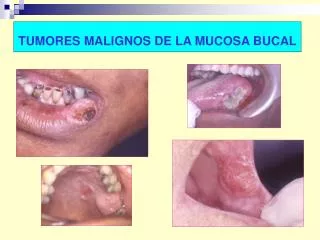
Diagnóstico diferencial • Hepatocarcinoma fibrolamelar • Mujeres jóvenes • AFP normal • Sin cirrosis • Metástasis • Adenocarcinomas • PAS/PASD • CDX-2 • TTF-1 • Carcinomas Hepatoides • Hep Par 1 • Ovario • Estómago • Vejiga Neoplasms of the liver. Goodman ZD. Department of Hepatic and Gastrointestinal Pathology, Armed Forces Institute of Pathology, Washington, DC, USA. Modern Pathology 2007 20, S49-s60
Biopsia con aguja • Separación trabéculas y fragmentación material • Ausencia de tejido conectivo y espacios porta (2X) • Células hepatocitarias con nucleolo, atipia, pérdida relación N/C • Pigmento biliar • Ausencia espacios porta • Pérdida de patrón de Reticulina • CD34 • Can CD34 discriminate between benign and malignant hepatocytic lesions in fine-needle aspirates and thin core biopsies?.De Boer WB et al. Division of Tissue Pathology, The Western Australian Centre for Pathology and Medical Research, WA, Australia. Cancer. 2000 Oct 25;90(5):273-8. • Distinction of hepatocellular carcinoma from benign hepatic mimickers using Glypican-3 and CD34 immunohistochemistry. Coston WM et al. Department of Pathology, City of Hope National Medical Center, Duarte, CA 91010, USA. Am J Surg Pathol. 2008 Mar;32(3):433-44.
Diagnóstico • Hepatocarcinoma bien diferenciado
Hepatocarcinoma5ª neoplasia en hombres y 8ª en mujeresTumor primario de hígado más frecuenteIncidencia geográfica variable Etiología Multifactorial Cirrosis 45-90% VHB VHC Anti-VHC antiC 15–80% de HCC Alcohol AFP Valores normales 40% de HCC S: 39-64% Esp: 76-91% VPP: 9-32% Marc derivados: PIVKAII AFP fracciones (L3) Alfafucosidasa Glipicano 3 Enfermedades Metabólicas Déficit alfa1 antitripsina Hemocromatosis Tirosinemia Porfiria Toxinas y fármacos Aflatoxina B1 Diabetes Descartar metástasis de tumor primario en hígado no cirrótico
Tratamiento • Cirugía: tumores pequeño tamaño, buena función hepática, sin HTPortal y asintomáticos • Trasplante: múltiples tumores pequeño tamaño si cirrosis y compensación • Ablación percutánea: Irresecables o no candidatos a trasplante. HTPortal, tumores únicos o 3 nódulos < 3cm • Radiofrecuencia • Etanol • Embolización arterial: lipiodol y quimioterápicos • Buen estado funcional y tumores grandes multinodulares • Tratamiento sintomático • Estadio avanzado • Sorafenib (Inhibidor tirosinquinasa) estudios fase III