Download
1 / 15

E N D
SKIN CANCER DETECTION USING CNN/GAN FINAL YEAR PROJECT BTI Prasant Poudel 1BH19CS069 Jitendra Kohar 1BH19CS034 Snehal Karki 1BH19CS103 Sanjay Malla 1BH19CS094
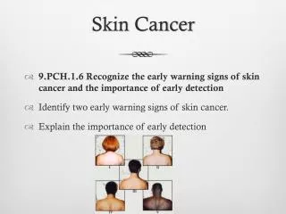
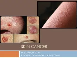
SKIN CANCER Skin cancer is the most commonly diagnosed cancer. Skin cancers are either non-melanoma or melanoma. Early detection and treatment can often lead to a highly favourable diagnosis. The visibility of the skin lesions increase the likelihood of early detection and diagnosis.
TYPE OF SKIN CANCER The most common types of skin cancer are: Basal Cell Carcinoma (BCC) Squamous cell carcinoma Malignant Melanoma
ABSTRACT Skin cancer is the most common form of cancer affecting humans. Melanoma is the most dangerous type of skin cancer; and early diagnosis is extremely vital in curing the disease. So far, the human knowledge in this field is very limited, thus, developing a mechanism capable of identifying the disease early on can save lives, reduce intervention and cut unnecessary costs. In this paper, the researchers developed a new learning technique to classify skin lesions, with the purpose of observing and identifying the presence of melanoma. This new technique is based on a convolutional neural network solution with multiple configurations; where the researchers employed an International Skin Imaging Collaboration (ISIC) dataset. Optimal results are achieved through a convolutional neural network composed of 14 layers.
INTRODUCTION Skin cancer is one of the most common cancers worldwide. There are several types of skin cancer. Basal cell carcinoma (BCC) and squamous cell carcinoma (SCC) are by far the most prevalent forms of skin cancer. BSS and SCC start in the epidermis, i.e. the outer layer of the skin, and since they are typically caused by sun exposure, they often develop in sun-exposed areas such as the face, ears, neck, head, arms, and hands. BCC rarely spread to other parts of the body, but SCC may spread to nearby organs or lymph nodes. Since BCC and SCC originate in keratinocytes (the most common type of skin cells), BCC and SCC are sometimes called keratinocyte cancers. Actinic Keratoses (Solar Keratoses) and Intraepithelial Carcinoma (Bowen’s disease) which are referred to in this paper as AKIEC, are common non-invasive lesions that are precursors of SCC. If untreated, they may progress to invasive SCC. Yet, many of the dermoscopic features and algorithms are complex, confusing and difficult to discover. As such, many dermatologists do not use dermoscopic tools correctly or accurately; which can result in clinical care risk. Completely different imaging techniques, such as multi-spectral imaging and confocal research, are forced to deal with the problems encountered while detecting melanoma tumors. and techniques needs to be trained in these imaging modalities. It is proven that dermoscopic examination by trained and experienced doctors yields higher sensitivity and specificity in the diagnosis of skin lesion. Therefore, the automated technique for robust analysis of dermoscopic dataset wibe extremely helpful for physicians
CONTINUE.. In the last few years, deep convolutional neural networks (CNN) become very popular in feature learning and object classification. Additionally, it has been widely used in medicine dataset, such as skin lesion analysis. The fact that, totally different features get detected at the various convolutional layers, permit the network to be handled automatically; therefore, resolving the difficulties of feature detection work present in convolutional pattern analysis techniques. Deep learning, particularly the convolutional neural network (CNN), has been widely applied to unravel several issues in computer vision. Varied CNN primarily based models developed for object classification and detection, such as Alex Net [8], VGG [9], GoogleNet [10], or ResNet [11]; are trained via the large image database ImageNet and have over 1000 training images for each training session.
CNN MODEL The idea is to develop a simple CNN(Convolutional Neural Network) model from scratch, and evaluate the performance to set a baseline. The following steps to improve the model are: Data augmentation: Rotations, noising, scaling to avoid overfitting Transferred Learning: Using a pre-trained network construct some additional layer at the end to fine tuning our model. Full training and Evaluation
EXISTING SYSTEM At present, to check skin malignancy of a patient, he needs to experience singular screening by a dermatologist so as to recognize whether they have skin disease or not. This framework helps dermatologist to process various cases a lot quicker than expected.There are a number of symptomatic checklist have been established. ABCDE is one of the checklists, such as - Asymmetry(A) - One portion of the affected cell that has turned into a tumor does not coordinate the other half. Wattage for this factor is 1.3. Border(B)-The edges/the fringe of the tainted cells wind up battered, scored, obscured. For this corresponding factor, the wattage is 0.1. Color(C)-Shade isn’t uniform. Shades of tan or dark colored spots on skin and dark are available. Dashes of red, white and blue add to the repulsive appearance. The wattage for this factor is 0.5. Diameter(D)- The cell width ends up more noteworthy than 6mm and over. Evolution(E)-Previously mentioned changes or advancements show MalignantMelanoma
PROPOSED SYSTEM The labeled images "benign" and "malignant" were used in this system. The images labeled as "other and unknown" were not used since the images in those groups could not be diagnosed. Images were put into dataset relying upon their analysis mark which has been extracted from the metadata of the pictures. The dataset has been organized in to two classes one containing all the dangerous dermoscopic pictures and other containing favorable dermoscopic pictures. The images from ISIC dermoscopic archive have been choosen randomly for the experimental section. In our proposed system, there exist three layers. First layer is the input layer where the datasets are trained on. Input layer collects data that are delivering and adding some weight with it that goes to hidden layers. The neurons of hidden layer separate the features from the data to ?ndout a pattern. The pattern is then used as basis to output layersthat selects to appropriate classes. Finally, binary classfication are used which appropriately select class 1 and class 0. For our case, class 0 means no harmful cells are present and class 1 means having malignant cancerous cells. How our system are implemented using convolutional neural network are depicted in Figure 1.
Figure 1. Convolutional neural network with its multiplelayers Figure 2. Flow chart for the system using convolutional neural network
LITERATURE SURVAY After carrying a survey on around 10 research paper, we can draw the inference that. There is still a lot of scope for rearch in the field of image processing for skin cancer detection and it can be furthermore used to reduce the number of deaths caused by melanoma and other kinds of cancer. Image-based computer aided diagnosis systems have much significant potential for screening and early detection of maligant melanoma. We reviewed the state of the art in these systems and then examine the cureent practices, problems, and prospects of image acquisition, pre_processing, segmentation, feature extraction and selection, and classification of demoscopic images. The incidence of skin cancer incidents has been drastically elevating day-to-day.Skin cancer in early stage could be cured easily by simple procedures or techniques but medication. So there is a need to detect and treat disease at an early stage.Overall, 4% of the cancer casses are melanoma.
UV-A and B are mainly responsible for skin cancer. Outdoor workers are generally more prone to skin cancer because they get easily exposed to skin cancers. So, precautionary measures like an application of sunscreen lotions need to be done. It can be treated at initial stages, as the duration is extended, the chances for treating skin cancer gets hastened. New molecular therapeutic approaches for skin cancer include several medications like cryosurgery, immune modulation with imiquimod, 5-FU, photodynamic therapy, etc. To mographic imaging of any soft tissue like the skin has a potential role in cancer detection. The penetration of infrared wavelengths makes a confocal approach based on laser feedback interferometry feasible. Experimental results were in agreement with numerical simulations and structural changes were evident which would permit discrimination of healthy tissue and tumor. Furthermore, cancer type discrimination was also able to be visualized using this imaging technique.
CONCLUTION The aim of this project is to determine the accurate prediction of skin cancer and also to classify the skin cancer as malignant or non-malignant melanoma. To do so, some pre- processing steps were carried out which followed Hair removal, shadow removal, glare removal and also segmentation. SVM and Deep Neural networks will be used to classify. classifier will be trained to learn the features and finally used to classify. The novelty of the present methodology is that it should do the detection in very quick time hence aiding the technicians to perfect their diagnostic skills. The dataset used is from the available ISIC (International Skin Image Collaboration) dataset, hence any dataset can be used to find the efficiency.