Download

1 / 48
570 likes | 905 Views
Electromyography (EMG). YU Yanqin, PhD Department of Physiology Zhejiang University School of Medicine. [Purpose]. 1. To explore the electrical activity of skeletal muscle by recording an electromyogram (EMG) from a volunteer.

E N D
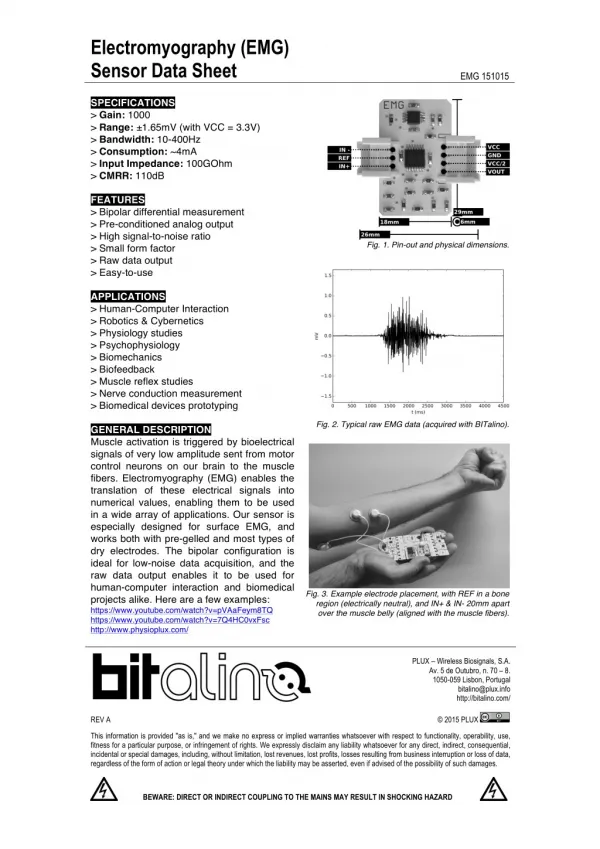
Electromyography (EMG) YU Yanqin, PhD Department of Physiology Zhejiang University School of Medicine
[Purpose] • 1. To explore the electrical activity of skeletal muscle by recording an electromyogram (EMG) from a volunteer. • 2. To examine the EMG of evoked muscle action and attempt to measure nerve conduction velocity.
[Principle] • The electrical signal recorded from a contracting muscle is called an electromyogram (EMG).
Medical uses of EMG • EMG is used as a diagnostics tool for identifying neuromuscular diseases, or as a research tool for studying kinesiology, and disorders of motor control. • EMG signals are also used as a control signal for prosthetic devices such as prosthetic hands, arms, and lower limbs.
Neuromuscular transmission A skeletal muscle fiber is innervated by a branch of a motor axon. Under normal circumstances, a neuronal action potential activates all of the muscle fibers innervated by the motor neuron. This activation process involves an action potential and a contraction of the muscle fibers. 1. Neuromuscular junction (NMJ) 2. Excitation-contraction coupling
Needle EMG and NCSs are typically indicated when there is pain in the limbs, weakness from spinal nerve compression, or concern about some other neurologic injury or disorder. NCS:nerve conduction studies
Like the electrocardiogram (ECG), EMG can be detected by electrodes placed on the skin. • A voluntary muscle contraction is produced by one or more action potentials in many muscle fibers (compound AP).
You will record EMG signals produced by electrical stimulation of a motor nerve supplying a muscle. • The abductor pollicis brevis muscle is a member of the thenar muscle group on the palmar surface of the hand. • The motor nerve to this muscle (the median nerve) is easy to stimulate at the wrist and elbow (Figure 1).
[Experimental method & procedure] • Select a volunteer for the experiment and have that person remove any watch, jewelry from their wrists. • Firmly attach the dry earth strap around the wrist of the volunteer. • Swab the skin with 75% alcohol in each area where electrodes will be placed on the volunteer. Put a small amount of electrode cream on the electrode surface. • With a ballpoint pen, lightly mark two small crosses on the skin overlying the abductor pollicis brevis muscle, in the position for the recording electrodes shown in Figure 2. The crosses should be 2–3 cm apart.
Figure 2. Electrode connections for recording from the abductor pollicis brevis muscle, and stimulation of the median nerve at the wrist and elbow.
Obtain two new disposable EMG electrodes and trim the adhesive pad slightly so they will fit as shown in Figure 2. Place a very small amount of electrode cream on the gelled surface of the electrode and attach them onto the skin over the crosses you marked. Use adhesive tape to fasten the electrode wires immediately adjacent to the skin electrodes. • Place a small amount of electrode cream on the two pads of the stimulating bar. • Place the bar stimulus electrode over the volunteer’s median nerve at the wrist. • The bar stimulus electrode should lie along the axis of the arm, with the leads pointing towards the hand. • Set the stimulator switch to the “ON” position.
Experiment procedure: • 1. Increase the pulse current to 10 mA. Apply a stimulus and record the response. Adjust the electrode to find the best position for stimulation. • 2. Increase the amplitude up to 20 mA in 2 mA increments, and record the response to a stimulus at each amplitude. The responses should increase with increasing stimuli until a maximal response is reached, after which increasing the stimulus does not further increase the response amplitude.
3. Save your file, remove the bar stimulus electrode and mark the spot in the pressure imprints on the skin. • 4. Then change to stimulate the median nerve in another position. • 5. Calculate the conduction velocity.
conduction velocity S 1- 2 S 1- 2 Δt υ= Δt
[Discussion] • 1. List the physiological events that occur between delivery of the electrical stimulus and the muscle contraction. • 2. What are the medical uses of EMG?
Electroencephalography (EEG) YU Yanqin, PhD Department of Physiology Zhejiang University School of Medicine
[Purpose] • To provide an introduction to the electroencephalogram. • To explore the electrical activity of the brain. • To examine the effects of visual activity on alpha waves.
[Principle] • The cerebral cortex contains large numbers of neurons. • Activity of these neurons is to some extent synchronized in regular firing rhythms (‘brain waves’). • Electrodes placed in pairs on the scalp can pick up variations in electrical potential that derive from this underlying cortical activity. • EEG signals are affected by the state of arousal of the cerebral cortex, and show characteristic changes in different stages of sleep. • Electroencephalography is also used in the diagnosis of epilepsies and the diagnosis of brain death.
EEG recording is technically difficult, mainly because of the small size of the voltage signals (typically 50 µV peak-to-peak). • The signals are small because the recording electrodes are separated from the brain’s surface by the scalp, the skull and a layer of cerebrospinal fluid. • A specially designed amplifier, such as the Bio Amplifier, is essential. • It is also important to use electrodes made of the right material, and to connect them properly. • Even with these precautions, recordings may be spoiled by a range of unwanted interfering influences, known as ‘artifacts’. • In clinical EEG, it is usual to record many channels of activity from multiple recording electrodes placed in an array over the head.
Medical uses of EEG • EEG is most often used to diagnose epilepsy, which causes abnormalities in EEG readings. • It is also used to diagnose sleep disorders, coma, encephalopathies, and brain death. • EEG used to be a first-line method of diagnosis for tumors, stroke and other focal brain disorders, but this use has decreased with the advent of high-resolution anatomical imaging techniques such as magnetic resonance imaging (MRI) and computed tomography (CT). • Despite limited spatial resolution, EEG continues to be a valuable tool for research and diagnosis, especially when millisecond-range temporal resolution (not possible with CT or MRI) is required.
EEG and brain death Brain death: No EEG signal for 12h
Measurement of EEG The international 10-20 system (Jasper, 1958)
Measurement of EEG in different brain areas Symmetrical in both hemisphere
[Experimental method & procedure] Figure 2. The equipment setup for this experiment, showing the placement of EEG flat electrodes on the head of the subject.
1. Subject preparation: The volunteer will need a place to lie on their back. The supine position reduces interference and results in better measurements. • 2. Tie the cap firmly around the head (Figure 2). Lightly abrade the skin over the mark with an abrasive pad/gel. Connect the electrodes for EEG measurement. If you are using electrode cream, squeeze about two drops into the side of the sponge of the electrode. • 3. Check that all electrodes are properly connected to the volunteer and the Bio Amp cable before proceeding. • 4. Starting the software: You are now ready to begin the exercises.
5. To examine alpha waves (alpha rhythm) in the EEG, and the effect of opening the eyes: Ensure that the subject is relaxed, lying quietly and has both eyes closed.
[Discussion] • 1. How do the brain waves correlate with the different stages of sleep?
Reflexes YU Yanqin, PhD Department of Physiology Zhejiang University School of Medicine
[Purpose] • 1. To investigate your reflexes in response to a variety of stimuli. 2. To examine some simple and complex reflexes from a volunteer.
[Principle] • Reflex: • In our day-to-day lives, we detect changes in the environment and react appropriately. Reflexes are examples of this type of stimulus-response reaction. A loud sound or something flying at your eye makes you blink, while a tap on the tendon under the kneecap produces the knee jerk, or myotactic, reflex. • Reflex arc: • An external stimulus is detected by sensory neurons, which send the information to the central nervous system, where it is processed. If a motor response is initiated, it usually involves a series of action potentials that produce a muscle contraction and a movement of one or more parts of the body.
The stretch reflex (myotatic reflex) • is a muscle contraction in response to stretching within the muscle. • It is a monosynaptic reflex which provides automatic regulation of skeletal muscle length.
Reflex arc of myotatic reflex • When a muscle lengthens, the muscle spindle is stretched and its nerve activity increases. This increases alpha motor neuron activity, causing the muscle fibers to contract and thus resist the stretching. • A secondary set of neurons also causes the opposing muscle to relax. • The reflex functions to maintain the muscle at a constant length.
Figure 1. Cross-section of the spinal cord, showing the neuronal circuitry of the myotactic reflex.
[Experimental method & procedure] • 1. The quadriceps (patellar reflex, knee jerk) reflex: 1) This is most easily done with the patient seated, feet dangling over the edge of the exam table. If they cannot maintain this position, have them lie supine (i.e. on their backs).
2) Identify the patellar tendon, a thick, broad band of tissue extending down from the lower aspect of the patella (kneecap). If you are not certain where it is located, ask the patient to extend their knee. This causes the quadriceps (thigh muscles) to contract and makes the attached tendon more apparent. • 3) Strike the tendon directly with your reflex hammer. • 4) In the normal reflex, the lower leg will extend at the knee.
Purpose of testing (knee jerk) • The test itself assesses the nervous tissue between and including the L2 and L4 segments of the spinal cord. • The absence or decrease of this reflex is problematic, and known as Westphal's sign. This reflex may be diminished or absent in lower motor neuron lesions and during sleep. • On the other hand, multiple oscillation of the leg (pendular reflex) following the tap may be a sign of cerebellar diseases. • Exaggerated (brisk) deep tendon reflexes such as this can be found in upper motor neuron lesions, hyperthyroidism, anxiety or nervousness.
Triceps reflex • 1) This is most easily done with the patient seated. • 2) Identify the triceps tendon, a discrete, broad structure that can be palpated (and often seen) as it extends across the elbow to the body of the muscle, located on the back of the upper arm. If you are having trouble clearly identifying the tendon, ask the patient to extend their lower arm at the elbow while you observe and palpate in the appropriate region. • 3) The arm can be placed in either of 2 positions: Gently pull the arm out from the patient’s body, such that it roughly forms a right angle at the shoulder. The lower arm should dangle directly downward at the elbow. Or have the patient place their hands on their hips. Either of these techniques will allow the triceps to completely relax.
4) If you are certain as to the precise location of the tendon, strike this area directly with your hammer. Or place your index or middle finger firmly against the structure. Then strike your finger. • 5) Make sure that the triceps is uncovered, so that you can observe the response. • 6) The normal reflex will cause the lower arm to extend at the elbow and swing away from the body.
[Discussion] • 1. What is the reflex arc for the myotactic reflexes such as the quadriceps or triceps reflex? • 2. What is the response of the pupil when light is shone on it? • 3. What is the response of the pupil in the opposite eye? • 4. What happens to pupil diameter when the eye is focused for near vision?