Download
1 / 6
280 likes | 1.3k Views
Electromyography. Tatiana Steinwarz. What is it?.

E N D
Electromyography Tatiana Steinwarz
What is it? • Electromyography, or EMG, involves testing the electrical activity of muscles. Often, EMG testing is performed with another test that measures the conducting function of nerves. This is called a nerve conduction study. Because both tests are often performed at the same office visit and by the same personnel, the risks and procedures generally apply to both tests. • Muscular movement involves the action of muscles and nerves and needs an electrical current. This electrical current is much weaker than the one in your household wiring. • In some medical conditions the electrical activity of the muscles or nerves is not normal. Finding and describing these electrical properties in the muscle or nerve may help your doctor diagnose your condition. • EMG may aid with the diagnosis of nerve compression or injury (such as carpal tunnel syndrome), nerve root injury (such as sciatica), and with other problems of the muscles or nerves. Less common medical conditions include amyotrophic lateral sclerosis, myasthenia gravis, and muscular dystrophy.
Diagnosis of neuromuscular disorders with the use of an electromyograph.
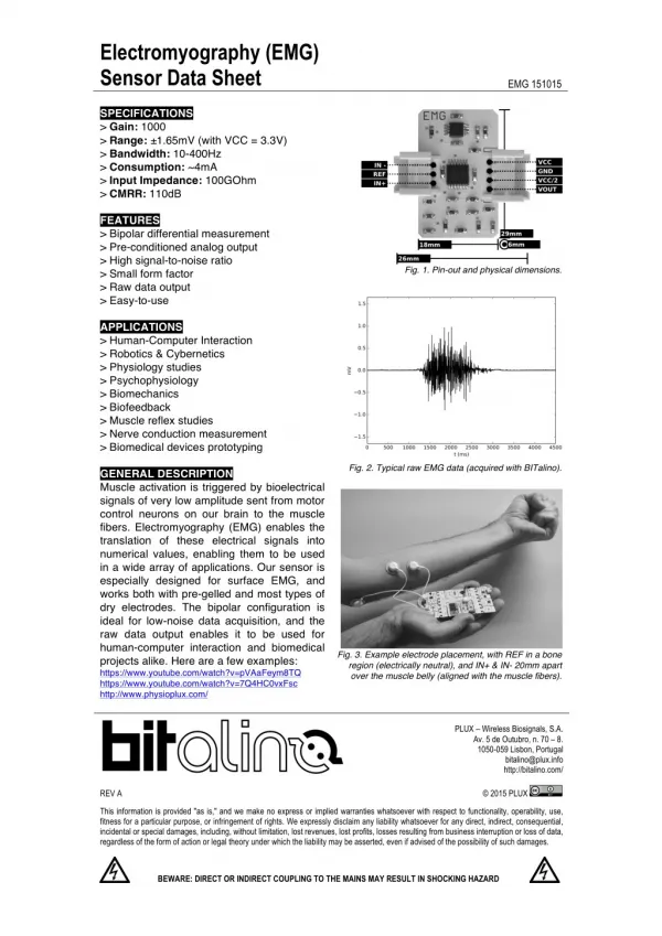
Procedure • To perform EMG, a tiny needle electrodes are inserted through the skin into the muscle tissue. Each electrode gives only an average picture of the activity of the selected muscle, because skeletal muscles are isolated and often large units. Several electrodes may need to be placed at various locations to obtain an accurate study. Then the patient is asked to contract the muscle. Each muscle fiber that contracts produces an action potential (a change in electrical potential), which is displayed on the electromyogram. The form of the action potential provides information about the ability of the muscle to respond when the nerves are stimulated. The size of the muscle fiber affects the rate (frequency) and size (amplitude) of the action potentials. • A nerve conduction velocity test is often done at the same time as an EMG. • Because of the needle electrodes, EMG may be somewhat painful to the patient, and the muscle may feel tender for a few days. There also exists "needleless EMG"—an EMG performed using surface electrodes—though it gives much less accurate results with a higher level of disturbance from the surrounding environment.
Why it is done? • Results from an EMG can help diagnose a condition that interferes with muscle contractions. These conditions include: • Diseases that affect the muscle, such as muscular dystrophies • Diseases that affect the connection between the nerve and the muscle (neuromuscular junction), such as myasthenia gravis • Diffuse nerve disorders that cause peripheral neuropathy • Disorders that affect the motor neurons (anterior horn cells) in the spinal cord, such as amyotrophic lateral sclerosis or a ruptured spinal disk
Overview • Definition: • Electromyography (EMG) is a test that checks the health of the muscles and the nerves that control the muscles. • Why it is performed: • EMG is most often used when people have symptoms of weakness and examination shows impaired muscle strength. It can help to differentiate primary muscle conditions from muscle weakness caused by neurologic disorders.