Download
1 / 34
411 likes | 1.03k Views
Electromyography (EMG). &. Motor Nerve Conduction Velocity. Dr Thouraya. Motor Unit. Consists of a motor neuron and all the muscle fibers it innervates When an action potential occurs in a motor neuron, all the Msl fibers in its MU are stimulated to contract.

E N D
Electromyography(EMG) & Motor Nerve Conduction Velocity Dr Thouraya
Motor Unit • Consists ofa motor neuron and all the muscle fibers it innervates • When an action potential occurs in a motor neuron, all the Msl fibers in its MU are stimulated to contract
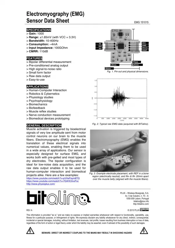
EMG is the recording of electrical activity of a Msl at rest & during contraction: (to evaluate the electrophysiology of a MU) • Activity is amplified and displayed on anoscilloscope. Instrument : Electromyograph Record: Electromyogram
A concentric needle Ede inserted into the belly of the Msl .
Needle EMG does not introduce any electrical stimulation instead it records the intrinsicelectrical activity of skeletal muscle fibers. • Normally a muscle is silent at rest after insertional activity has ceased.
Then the patient is asked to contract the Msl smoothly. • With muscle contraction, MUs are activated andMUAPsappear on the screen: • Motor unit potential : represents the summation of the potentials generated byµslfibersbelonging to theMU
Normal MUPs • Bi – Triphasic • Duration – 3 – 16 mSec. • Amplitude – 300μV – 5 mV
With increasing strength of contracto →recruitment of MUs →↑number & size of MUAPs • At full contraction separate MUAP will be indistinguishable resulting in a complete recruitment = interference pattern
Analysis The EMG is used to investigate both neuropathic and myopathic disorders (weakness,numbness,pain ) • The size, duration, frequency of the electrical signals generated by Msl cells help determine if there is damage to the Msl or to the nerve leading to that Msl.
Myopathy: progressive degeneration of skleletal muscle fibers Eg: Duchenne Muscular dystrophy
Neuropathy:Damage to the distal part of the nerve. peripheral neuropathy mainly affects feet & legs Most common etiologies: • Guillain Barré syndrome • Diabetes mellitus • Alcohol abuse
LMN lesions: interrupt the spinal reflex arc ( α motor N) →Partial or complete loss of voluntary contraction , muscle wasting,↓reflexes, fasciculation Example: Polyomyelitis
In neurogenic lesion or in active myositis, the following spontaneous activity is noted: • Positive sharp waves • Fibrillations • Giant motor unit potentials
Fibrillation potentials: Low amplitude, short duration potentials, correspond to the spontaneous discharge of adenervated single muscle fiberdue to denervato hypersensitivity to acetylcholine. Fine invisible,irregular contractions of individual muscle fibers.
Positive sharp waves Small fibrillation APs (50 to 100 µV, 5 to 10 msec duration) whose propagation is blocked at the level of the recording Ede
Fasciculation potentials spontaneous discharge of a MU at rest, can be seen and felt by the patients • Partial re-innervationof denervated muscle, by sprouting of the remaining nerve terminals, produces abnormally large, long polyphasic potentials (giant potential)
Myopathic alteration of the EMG: Polyphasia ,short duration ,reduced voltage of MUPs
Neuropathic alteration of the EMG: Polyphasia ,long duration ,high voltage of MUPs
Nerve Conduction studies A nerve conduction study (NCS) is a test commonly used to evaluate the function, especially the ability of electrical conduction, of the motor and sensory nerves of the human body.
Motor Nerve Conduction Study • Stimulato of mediannerve at two points until visible muscle contracto is seen and a reproducible Compound Muscle A P is recorded CMAP:summated potentials from all Motor Units in a muscle
distance l1- l2 (m/sec) • MNCV= l1 = latency at elbow. l2 = latency at wrist Distance between the two stimulating electrodes • abNl if < 40 m/sec
Normal values for conduction velocity • In arm • 50 to 70 m / sec. • In leg • 40 to 60 m / sec.
Conduction is faster in myelinated fibres. • Diseases which produce demyelinated peripheral nerves (diabetes,Gillain Barré)slow the conducto greatly(20-30 m/s).