Download

1 / 10
500 likes | 1.91k Views
Electromyography ( EMG ). Dr Malith Kumarasinghe MBBS (Colombo). Research Applications of Surface EMG. Indicator for muscle activation/deactivation Relationship of force/EMG signal Use of EMG signal as a fatigue index. Types of EMG. Electrode Categories Inserted

E N D
Electromyography (EMG) Dr Malith Kumarasinghe MBBS (Colombo)
Research Applications of Surface EMG • Indicator for muscle activation/deactivation • Relationship of force/EMG signal • Use of EMG signal as a fatigue index
Types of EMG • Electrode Categories • Inserted • Fine-wire (Intra-muscular) • Needle • Surface
Fine-wire Electrodes • Advantages • Extremely sensitive • Record single muscle activity • Access to deep musculature • Little cross-talk concern • Disadvantages • Requires medical personnel, certification • Repositioning nearly impossible • Detection area may not be representative of entire muscle
Surface Electrodes • Advantages • Quick, easy to apply • No medical supervision, required certification • Minimal discomfort • Disadvantages • Generally used only for superficial muscles • Cross-talk concerns • No standard electrode placement • May affect movement patterns of subject • Limitations with recording dynamic muscle activity
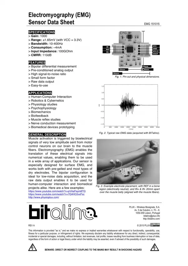
Characteristics of EMG Signal • Amplitude range: 0–10 mV (+5 to -5) prior to amplification • Useable energy: Range of 0 - 500 Hz • Dominant energy: 50 – 150 Hz
Electrode Placement • Away from tendon • Fewer, thinner muscle fibers • Closer to other muscle origins, insertions • More susceptible to cross-talk • Away from outer edge of muscle • Closer to other musculature • Orientation parallel to muscle fibers • More accurate conduction velocity • Increased probability of detecting same signal
Reference Electrode Placement(Ground) • As far away as possible from recording electrodes • Electrically neutral tissue • Bony prominence • Good electrical contact • Larger size • Good adhesive properties