Download
1 / 2
40 likes | 262 Views
EMG enables the translation of these electrical signals into numerical values, enabling them to used in a wide array of applications.

E N D
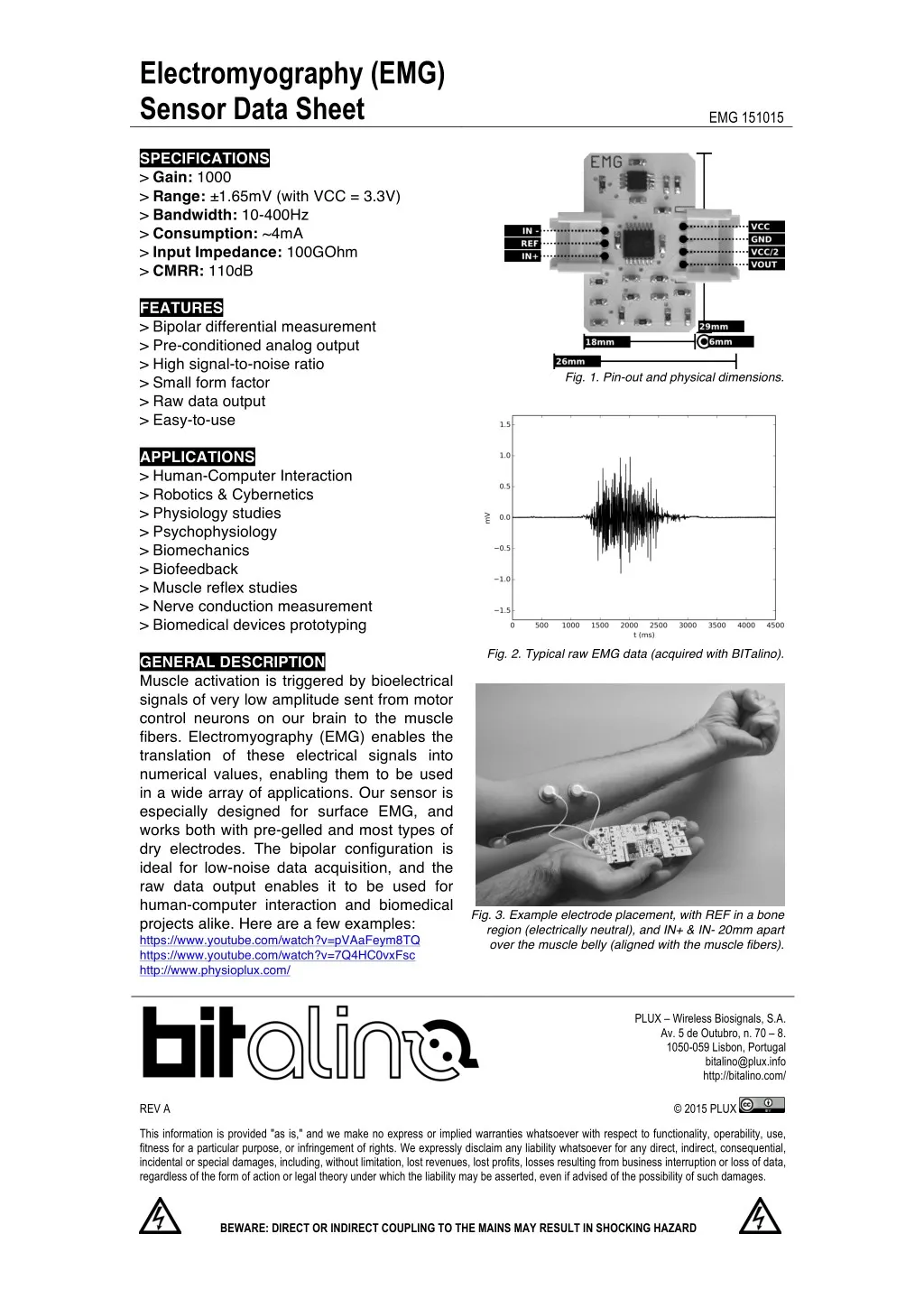
Electromyography (EMG) Sensor Data Sheet SPECIFICATIONS > Gain: 1000 > Range: ±1.65mV (with VCC = 3.3V) > Bandwidth: 10-400Hz > Consumption: ~4mA > Input Impedance: 100GOhm > CMRR: 110dB FEATURES > Bipolar differential measurement > Pre-conditioned analog output > High signal-to-noise ratio > Small form factor > Raw data output > Easy-to-use APPLICATIONS > Human-Computer Interaction > Robotics & Cybernetics > Physiology studies > Psychophysiology > Biomechanics > Biofeedback > Muscle reflex studies > Nerve conduction measurement > Biomedical devices prototyping GENERAL DESCRIPTION Muscle activation is triggered by bioelectrical signals of very low amplitude sent from motor control neurons on our brain to the muscle fibers. Electromyography (EMG) enables the translation of these electrical signals into numerical values, enabling them to be used in a wide array of applications. Our sensor is especially designed for surface EMG, and works both with pre-gelled and most types of dry electrodes. The bipolar configuration is ideal for low-noise data acquisition, and the raw data output enables it to be used for human-computer interaction and biomedical projects alike. Here are a few examples: https://www.youtube.com/watch?v=pVAaFeym8TQ https://www.youtube.com/watch?v=7Q4HC0vxFsc http://www.physioplux.com/ EMG 151015 Fig. 1. Pin-out and physical dimensions. Fig. 2. Typical raw EMG data (acquired with BITalino). Fig. 3. Example electrode placement, with REF in a bone region (electrically neutral), and IN+ & IN- 20mm apart over the muscle belly (aligned with the muscle fibers). PLUX – Wireless Biosignals, S.A. Av. 5 de Outubro, n. 70 – 8. 1050-059 Lisbon, Portugal bitalino@plux.info http://bitalino.com/ REV A This information is provided "as is," and we make no express or implied warranties whatsoever with respect to functionality, operability, use, fitness for a particular purpose, or infringement of rights. We expressly disclaim any liability whatsoever for any direct, indirect, consequential, incidental or special damages, including, without limitation, lost revenues, lost profits, losses resulting from business interruption or loss of data, regardless of the form of action or legal theory under which the liability may be asserted, even if advised of the possibility of such damages. © 2015 PLUX BEWARE: DIRECT OR INDIRECT COUPLING TO THE MAINS MAY RESULT IN SHOCKING HAZARD
Electromyography (EMG) Sensor Data Sheet TRANSFER FUNCTION [-1.65!", 1.65!"] !"# 2! −1 !!"# !"# !" = !"# ! .1000 !"" = 3.3! (operating voltage) !!"#= 1000 (sensor gain) !"# ! – EMG value in Volt (!) !"# !" – EMG value in millivolt (!") !"# – Value sampled from the channel ! – Number of bits of the channel1 2.!"" !"# ! = 1 The number of bits for each channel depends on the resolution of the Analog-to-Digital Converter (ADC); in BITalino the first four channels are sampled using 10-bit resolution (! = 10), while the last two are sampled using 6-bit (! = 6). PAGE 2 OF 2