Download
1 / 31
390 likes | 1.1k Views
Scratch for Arduino (S4A) and the Electromyography (EMG) Sensor. Mitchell Neilsen INSIGHT Summer Institute 2014. Cyber-Physical Systems (CPS). CPS Future Potential.

E N D
Scratch for Arduino (S4A) and the Electromyography (EMG) Sensor Mitchell Neilsen INSIGHT Summer Institute 2014
CPS Future Potential • Ongoing advances in science and engineering will improve the link between computational and physical elements, dramatically increasing the adaptability, autonomy, efficiency, functionality, reliability, safety, and usability of cyber-physical systems. • This will broaden the potential of cyber-physical systems in several dimensions, including: • intervention (e.g., collision avoidance); • precision (e.g., robotic surgery and nano-level manufacturing); • operation in dangerous or inaccessible environments (e.g., search and rescue, firefighting, and deep-sea exploration) • coordination (e.g., air traffic control, war fighting); • efficiency (e.g., zero-net energy buildings); and • augmentation of human capabilities (e.g., healthcare monitoring, prosthetics, etc.).[2]
Electromyography (EMG) • Electromyography (EMG) is a technique for evaluating and recording the electrical activity produced by skeletal muscles – from wikipedia • Study that deals with the detection, analysis, and use of electrical signals that emanate from contracting muscles – from “The physiology background of EMG” by LidaMademli
Skeletal Muscle Organization Muscle consists of: • Muscle fascicles (bundles of muscle fibres) • Muscle fascicles are wrapped by perimysium • Muscle fascicles consist of • Muscle fibres (muscle cell) • Muscle fibres are wrapped by endomysium • The muscle fibreis what contracts
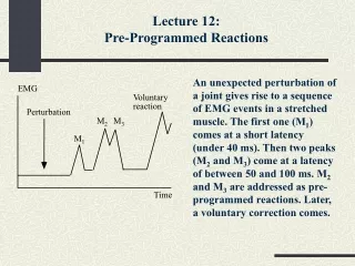
Motoneuron • How the electrical stimulus travels down the motoneuronto innervate (activate) the muscle fibre. • The change in polarity travels down the neuron (action potential) • Neurotransmitter(acetylcholine) is released from terminal end
Muscle at Rest Resting potential of muscle = ~ -90mV (Purves et al 2001) • In the absence of an impulse, the inside is electrically negative and the outside is positive
Muscle and Nerve The Motor Unit (MU) • One muscle may have many motor units of different fibretypes (slow or fast twitch) • One motor unit can have from 5 to few thousands muscle fibres. • The motor unit is the brain’s smallest functional unit of force development control.
EMG Signal Capture • Differential amplifier • Input from two different points of the muscle • Close (usually 1-2cm) • Electrode alignment with the direction of muscle fibres implies increased probability of detecting same signal • Subtracts the two inputs • Amplifies the difference • Optionally, rectify and smooth signal
Physiology of the EMG • Above the innervation zone, electrode 7 small amplitude. • Above the myotendinousjunction, more tendon tissue, electrodes 14 and 15 small amplitudes. • Others radiate out from electrode 7. electrode
Types of EMG Sensors sEMG = surface EMG
Characteristics of EMG Signal • Amplitude range: 0 - 10 mV (+5 to -5) prior to amplification • EMG frequency: range of 10 - 500 Hz • Dominant energy: 50 - 150 Hz • Peak in the neighborhood of 80 - 100Hz
EMG Frequency • Motor Units • Slow twitch: 75 - 125 Hz (twitches/sec) • Fast twitch: 125 - 250 Hz
Most Usual Parameters in Biomechanics or Physiology • Time domain: • RMS or average of rectified EMG • Frequency domain: • FFT analysis (spectral analysis) • Power density • Mean power frequency • Median power frequency
Potential Use of EMG • It is possible that the EMG signal during a specific movement will demonstrate inter-individual differences. This can be used in user authentication systems (ACTIBIO).
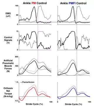
Potential Use of EMG • The muscle activity recorded using sEMG can be useful as an input signal to the system which can control devices such as keyboard, mouse or computer. (Arjunan, et al., 2007) • “The strength of sEMG is a good measure of the strength of contraction of muscle”. (Arjunan, et al., 2007) • Not always true! E.g., EMG During Fatigue
Muscle Sensor • Advancer Technologies Muscle Sensor v3: http://www.advancertechnologies.com/p/muscle-sensor-v3.html
Rectified and Smoothed Signal • These sensors do not output a RAW EMG signal, but rather an amplified, rectified, and smoothed signal the can be used directly with a microcontroller’s analog-to-digital converter. • This difference can be illustrated using a simple sine wave as an example.
Connect Sensors • See http://www.youtube.com/watch?v=VnrsWdA6dzE&feature=player_embedded
Connect to Arduino • Simple Scratch4Arduino Example
Now, put the two together • If the muscle activity reaches a sufficient level, then turn on the external device… • If the muscle activity drops below a certain level, then turn the external device off…
Conductive Fabric Electrode Sleeve • Cut out three rectangular strips of the conductive fabric. Two of the strips should be W 5/8" x L 1 3/4". The third strip should be W 5/8" x L 2". • Take the forearm sleeve, turn it inside out, and put it on the opposite arm that it is intended to go on. • Using fabric pins, pin the two shorter strips on your forearm muscle such that one is in the middle of the muscle body and the other is about an inch apart. Pin the third strip along the back side of your forearm (on the bony part). Check out the pictures to see how to orient the strips. • Carefully take the sleeve off and you're ready to sew.
Conductive Fabric Material • "This medical grade Silver plated 76% Nylon, 24% elastic fiber fabric offers the unique ability to stretch in both directions. • Can be used as an antibacterial wound dressing (note: our material is not sterile) but it also makes a great material for electrode contacts, stretchy hats, socks, gloves, or other garments. • Highly conductive, and conductivity increases as it stretches in one direction, and decreases as it stretches in the other direction. • Silver coating is 99.9% pure. Silver/gray color. “ • lessemf.com
S4A Download • http://s4a.cat/