Download
1 / 49
490 likes | 502 Views
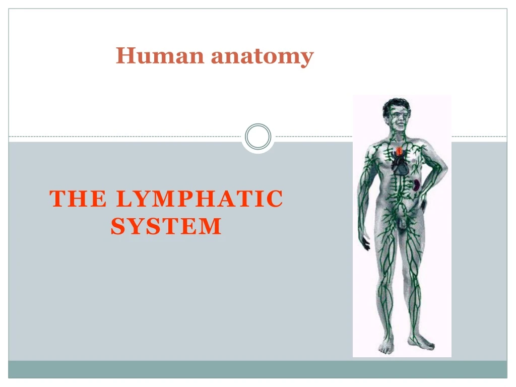
Human anatomy. THE LYMPHATIC SYSTEM. The Lymphatic system, Is a subsystem of the circulatory system that consists of a complex network of vessels, tissues, and organs. This network of lymphatic vessels carry a clear fluid called lymph in one direction t owards the heart.

E N D
Human anatomy THE LYMPHATIC SYSTEM
The Lymphatic system, Is a subsystem of the circulatory system that consists of a complex network of vessels, tissues, and organs. This network of lymphatic vessels carry a clear fluid called lymph in one direction towards the heart. The lymphatic system helps maintain Fluid balance in the body by collecting excess fluid and particulate matter from tissues and depositing them in the bloodstream. It also helps defend the body against infection by supplying disease-fighting cells called Lymphocytes. Definition
LYMPAHATIC SYSTEM • Lymphatic comes from the Latin word lymphaticus, meaning "connected to water," as lymph is clear. • This network of vessels & lymph nodes which are located in all major tissues of body. • Lymphatic system is absent in CNS, Cornea, Superficial layer of skin, Bones, Alveoli of lung. • CONSIST OF • Lymph • Lymphatic Channels • Lymph Nodes • Lymph Organs Capillaries Vessels Ducts
When circulating blood reaches the capillaries Part of its fluid passes intosurrounding tissues As a Tissue fluid Most of the fluid re-enters the capillaries at their venous ends Some of it return to the circulation through a separate system Lymphatic vessels/ Lymphatics
The lymph system is not a closed system The circulatory system processes an average of 20 litres of blood per day through capillary filtration which removes plasma while leaving the blood cells. Roughly 17 litres of the filtered plasma actually get reabsorbed directly into the blood vessels, while the remaining 3 litres are left behind in the interstitial fluid. The primary function of the lymph system is to provide an accessory route for these excess 3 litres per day to get returned to the blood. Therefore , The Lymph is essentially recycled blood plasma.
The lymphatic functions It is responsible for the removal of interstitial fluid from tissues It absorbs and transports fatty acids and fats from the digestive system It transports white blood cells to and from the lymph nodes into the bones The lymph transports antigen-presenting cells (APCs), such as dendritic cells, to the lymph nodes where an immune response is stimulated.
The LYMPHATIC SYSTEMS consists of: 1. Lymphatic vessels 2. Lymphoid tissues and lymphoid organs
CONTENTS • Development of lymphatic system • Lymphatic system • Lymph • Lymphatic Channels
Develop at the end of 5th wk of embryonic life Lymphatic vessels develop from lymph sacs which arise from developing veins and are derived from mesoderm 1st lymph sac to appear are the paired jugular lymph sacs at junction of internal jugular & subclavian veins DEVELOPMENT
JUGULAR LYMPH SACS • Retains one connection with its Jugular vein • Spreads lymphatic capillary plexuses to Thorax , upper limbs, head & neck. • The Left one develops into superior portion of thoracic duct. RETROPERITONEAL LYMPH SAC • It is unpaired and develops from primitive vena cava & mesonephric veins. • Spreads capillary plexuses & lymphatic vessels to abdominal viscera & diaphragm. • Develops connections with cisterna chyli & loses connections with neighboring veins
CISTERNA CHYLI • develops inferior to diaphragm on post abdominal wall. • gives rise to inferior portion of thoracic duct. POSTERIOR LYMPH SACS • Develops from iliac veins. • Gives capillary plexuses & lymphatic vessels to abdominal wall , pelvic region & lower limbs. • Join cisterna chyli & loose connections with adjacent veins
Lymph vessels grow out from the lymph sacs, along the major veins. • Except for the upper portion of the cisterna chyli, which persists, the lymph sacs are transformed into groups of lymph nodes during early fetal life, at about 3 months.
THE LYMPH • Transudative fluid. • Transparent & slightly yellowish liquid. • Alkaline in nature. • Derived from tissue fluid. • When blood passes through tissues 9/10 of fluid - venous end 1/10 of fluid - lymph capillaries • “CHYLE” - Lymph from small intestine.
COMPOSITION OF LYMPH 96% water 4% solids PROTEINS : 2 to 6 % of solids. Depending upon the part of body from which it is collected Albumin, globulin, clotting factors (fibrinogen, prothrombin) , all antibodies and enzymes. LIPIDS : 5-15 % - Mainly chylomicrons and lipoproteins. CARBOHYDRATES : Sugar - 132 mg per 100 ml (Mainly glucose). Non protein nitrogenous substances : Urea, A.A & Creatinine. ELECTROLYTES : Sodium, calcium, potassium, Chloride & bicarbonate. CELLULAR CONTENT : Mainly lymphocytes 1000-2000 per cu mm
lymphatic capillaries Are absent from bone, bone marrow and from the entire central nervous system. • The excess in nervous tissue fluid drains through the into the cerebrospinal fluid. • The cerebrospinal fluid then returns this tissue fluid to the blood through the superior sagittal sinus. • lymphatic capillaries, called lacteals, has a unique function. • Located in the villi of the mucosa of the small intestine, lacteals absorb digested fats from the intestine, which causes the lymph draining from the digestive viscera to become milky white. • This fatty lymph is called chyle, and, like all lymph, it is carried into the bloodstream.
LYMPHATIC CAPILLARIES • They are lined by a single layer of endothelial cells. • These are attached to C.T by anchoring filaments. • The edge of one endothelial cell overlaps the adjacent cell. • Overlapping edge is free to flap inward minute valve. • Permits passage of high molecular weight substance.
Flow of Lymph • Lymph takes the following route from the tissues back to the bloodstream: • lymphatic capillaries -> collecting vessels -> six lymphatic trunks -> two collecting ducts -> subclavian veins. • Thus, there is a continual recycling of fluid from blood to tissue fluid to lymph and back to the blood Anatomy and physiology - The unity of form and function (Saladin K. - 2003 - 3rd ed. - McGraw-Hill)
LYMPHATIC VESSELS • Lymph capillaries merge to form lymphatic vessels. • Resemble veins but …………. • Thin walls (Diameter - 0.2 – 0.3 mm) More valves (formed from folds of tunica intima) • Lymph Nodes are located at interval along its course Have 3 coats (Tunica intima, Tunica media, Tunica adventitia) BEADED in appearance ( due to semilunar valves). Collagenous fibers attaches the endothelium to the outer tissues ( fibrous sheath of muscle)
Lymph and Lymph Vessels • In contrast to the blood circulation, which uses the pumping of the heart to circulate its flow, lymph is propelled through the vessels primarily by the rhythmic contractions of tiny muscular units (lymphangions) which form the lymph collectors. • The lymphatic system has a slow rhythm, low velocity and low pressure. • Human body has twice as many lymph vessels and capillaries as you have blood vessels and capillaries!
Mechanism of lymphatic flow :- • Lymph flows under forces similar to those that govern venous return, except that the lymphatic system has no pump like the heart. • Lymph flows at even lower pressure and speed than venous blood; it is moved primarily by rhythmic contractions of the lymphatic vessels themselves, which contract when stretched by lymph. • The lymphatic vessels, like the veins, are also aided by a skeletal muscle pump that squeezes them and moves the lymph along. • Also like the medium veins, lymphatic vessels have valves that prevent lymph from flowing backward. • Since lymphatic vessels are often wrapped with an artery in a common sheath, arterial pulsation may also rhythmically squeeze the lymphatic vessels and contribute to lymph flow. • A thoracic (respiratory) pump aids the flow of lymph from the abdominal to the thoracic cavity as one inhales, just as it does in venous return. • Finally, at the point where the collecting ducts join the subclavian veins, the rapidly flowing bloodstream draws the lymph into it. • Considering these mechanisms of lymph flow, it should be apparent that physical exercise significantly increases the rate of lymphatic return.
How is fluid moved? - Contraction of skeletal muscles against lymphatic vessels - Smooth muscle contraction - Valves in lymphatic vessels
Lymphatic Trunks (Paired, except intestinal) • Lumbar • Intestinal Receives fatty lymph (chyle) absorbed through lacteals of intestines • Broncho-mediastinal • Subclavian • Jugular Drain into cisterna chyli
Lymphatic Trunks Left jugular trunk Right jugular trunk Left subclavian trunk Right subclavian trunk Right Broncho- Mediastinal Trunk Left Broncho- Mediastinal Trunk Thoracic Duct Cisterna Chyli Intestinal Trunk Right lumber Trunk Left lumber Trunk
THORACIC / LEFT LYMPHATIC DUCT • Thoracic duct (always present) (drain into left subclavian vein) • 38 – 45 cm long • Begins as a dilation called cisterna chyli anterior to 2nd lumber vertebra. • Main duct for return of lymph to blood • Receives lymph from left side of head, neck, Left upper limb, chest & entire body inferior to ribs • Joins the venous system at the junction of Left Sub clavian & Left internal jugular veins & drains lymph via Lt subclavian vein.
Origin, course, relations, and termination • Arises in the abdomen from cisterna chyli under cover ofdiaphragm. • Enters the thorax through the aortic opening. • It continues upward through the posterior mediastinum, on the left, first with the aortic arch and then with the left pleura. • It enters the root of the neck, where it arches laterally behind the left carotid sheath, to terminate in the upper end of the left innominate vein, in the angle of junction of the internal jugular and subclavian veins. • Chyle leak. • Virchowsor scalenenodes or signal nodes – supra clavicular nodes.
RIGHT LYMPHATIC DUCT • 1.2 cm long • 3 lymphatic trunks drain into Rt lymphatic duct • Rt Jugular trunk-drains Rt side of head & neck • Rt subclavian trunk-Rt upper limb • Rt bronchomediastinal trunk-Rt side of thorax, Rt lung, • Rt side of heart , & part of liver • Rt lymphatic duct joins the venous system at the junction of Rt Sub clavian & Rt internal jugular veins
Lymph Drainage of Lower Limb Superficial inguinal lymph nodes • One group parallel and below the inguinal ligament • Another group along the upper part of great saphenous vein • They drain all superficial structures in: • Buttocks, thigh, leg, foot • Anterior abdominal wall below the umbilicus. • External genitalia except testis • Perineum and lower parts of vagina and anal canal • Cornu of uterus
Lymph Drainage of Lower Limb Cont., Deepinguinal lymph nodes • They are located medial to femoral vein • They drain: lymphatic vessels that accompany femoral vessels and from popliteal fossa Glans penis and clitoris Efferent lymphatics from superficial inguinal lymph nodes • Efferent lymphatics from this group drain into external lymph nodes Superficial inguinal LN
Lymphatic Drainage of the Upper Limb • Superficial lymphatic vessels from the thumb, index and lateral side of the hand follow the cephalic vein to infraclavicular lymph nodes • Superficial lymphatic vessels from the medial side of the hand follow the basilic vein to supratrochlear lymph nodes then to the lateral group of axillary lymph nodes • Deep lymphatic vessels follow the arteries to the lateral group of axillary lymph nodes
Axillary Lymph Nodes • They drain lymph from entire upper limb, lateral part of breast, and superficial lymphtics of skin above the umblicus They include the following groups: • Anterior: deep to pectoralis major at lower border of pectoralis minor drain most of breast • Posterior: in front of subscapularis muscle • Lateral: along the axillary vein • Central: deep in the axilla • Infraclavicular • Apical: above the clavicle at the apex of axilla
Lymph Nodes of the Head & Neck Deep Cervical Lymph Nodes 7. Lateral jugular 8. Anterior jugular 9. Jugulodigastric Inferior Deep Cervical Lymph Nodes 10. Juguloomohyoid 11. Supraclavicular (scalene)
Deep Lymph Nodes 1. Submental 2. Submandibular (Submaxillary)
Anterior Cervical Lymph Nodes (Deep) 3. Prelaryngeal 4. Thyroid 5. Pretracheal 6. Paratracheal
Lymph Drainage of the Head and Neck Deep Cervical Lymph Nodes: • They drain lymph from the entire head and neck • Located along the whole length of internal jugular vein • They are two groups Upper group located at the angle between the lower border of mandible and anterior border of sternomastoid Lower group located in the angle between the clavicle and sternomastoid
Lymph Drainage of the Thorax Lymph nodes of the chest wall: • Parasternal • Intercostal • Diaphragmatic Lymph nodes of the mediastinum: • Nodes around bifurcation of trachea and main bronchi • Posterior mediastinal • Anterior mediastinal
Lymph Node Groups in Abdomen • They are related to the main arteries of the abdomen • Preaortic group: They are related to the main single branches of the aorta • Paraaortic group: They are related to the lateral branches of the aorta
Coeliac Nodes • Located around the stem of the coeliac trunk • They drain node groups related to the main arteries of the region: Left gastric Splenic Hepatic Gastroduodenal Gastroepiploic Pyloric pyloric LN
Superior and Inferior Mesenteric Lymph Nodes • Drain the small and large intestines • Drain the intestines via lymph nodes close to the intestinal wall and intermediate nodes in the mesentery
Lymphatic Drainage of the Pelvis • Lymph nodes along the external, internal, and common iliac vessels in addition to the sacral vessels • Lymph nodes between the layers of the broad ligament and in the fascial sheath of the rectum and the urinary bladder