Download
1 / 102
1.26k likes | 3.19k Views
Anatomy and diseases of the uvea. Anatomy:

E N D
Anatomy and diseases of the uvea Anatomy: Uvea is the vascular coat of eye ball and lies between the sclera and retina. Uvea is composed of three parts i.e. iris, ciliary body and choroid. These three portions are intimately connected and a disease of one part also affects the other portions though not necessarily to the same degree.
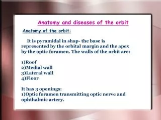
1) Iris: Iris is a delicate membrane placed in the anterior part of the eye ball and perforated in the center i.e. pupil. It arises from the middle of anterior surface of ciliary body. It is slightly pushed forward by the lens which gives it the appearance of a truncated cone, the apex of which has been cut off.
Anterior surface has two zones: 1) Papillary zone: is flat and has a dark border at the papillary margin , known as papillary ruff. Junction of papillary and ciliary zone is marked by a smooth ridge known as collaret. 2) Ciliary zone: towards the ciliary border has ciliary crypts. Histologically iris has got 5 layers (from before backward). 1- Endothelium: flat nucleated cells absent over the crypts probably to allow free movement of the aqueous in and out of the iris.
2- Vessel layer: consist of blood vessels lying in collagen fibers, chromotophores etc., 3- Muscular layer: two muscles: a) Sphincter pupillae- plain muscle developed from neuroectoderm- supplied by parasympathetic fibers coming through the 3rd nerve (relayed in ciliary ganglion). b) Dilator pupillae- also arise from neuroectoderm and supplied by cervical sympathetic fibers reaching the eye through long ciliary nerves (not relayed in ciliary ganglion).
4- Pigmented epithelium- two layers. The two layers are loosely attached to each other and there is a potential space between the 2 layers. In iridocyclitis there is adhesion of the iris to the lens. When a mydriatic is applied the posterior layer of pigment epithelium gets partly detached. This produces pigment deposition and pigmented patches on the anterior lens capsule (broken posterior synechiae).
5- Internal limiting membrane- fine and homogenous structure, not consistently present. Function of the iris: 1- Regulates the entry of light into the eye by changing the size of papillary aperture. 2- Cuts away the peripheral aberrations. 3-Absorption of aqueous also takes place from its surface.
2) Ciliary body: Ciliary body is a ring shaped structure placed more or less sagitally and extends from the ora-serrata to the scleral spur. It consists of ciliary processes and ciliary muscle. On longitudinal section it is triangular. It’s anterior surface or base is shortest. Iris is attached to the base. The outer side of triangle is adjacent to sclera and is formed by ciliary muscle.
The inner surface is directed towards the cavity of the eye ball and is divided into two portions anterior (pars plicata ) and posterior (pars plana). Ciliary muscle consists of: 1. Meridional fibers. 2. Circular fibers. 3. Radiating fibers.
Meridional fibers on contraction pull the suprachoroidea forwards and release the suspensory ligament allowing the lens to become more convex as in accommodation. The pars plicata has about 70 ciliary processes. They secrete aqueous. Functions of ciliary body: 1. Brings about accommodation. 2. Formation of aqueous. 3. Helps in drainage of aqueous at the angle of anterior chamber.
3) Choroid: Choroid is the analogue of pia-arachnoid of the brain and serves the same purpose of supplying nutrition to the neural portion of eye i.e. retina. Choroid is composed of five portions. 1. The outer most is SUPRACHOROIDEA a potential space between the choroid and sclera. This is lined by endothelium and traversed by fibrous trabeculae.
This space is utilized for the treatment of aphakic glaucoma in the operation of cyclodialysis. Deeper to it are three vascular layers. 2. Layer of LARGE BLOOD VESSELS is outer most. 3. Next comes MEDIUM SIZED BLOOD VESSELES. 4. and SMALL BLOOD VESSELES or CHOROIDO-CAPILLARIES.
Layer of choriocapillaries is the most important. It serves to provide nutrition to the outer layers of retina. The choriocapillaries are much wider than the capillaries elsewhere. Their diameter varies from 10 to 30 microns. 5. The innermost layer is avascular known as MEMBRANE of BRUCHS. This is composed of elastic and cuticular lamina and pigment epithelium of retina is intimately attached to it.
Classification: A) Depending upon the site of inflammation: 1) Anterior uveities - iritis. - cyclitis. - iridocyclitis. As iris and ciliary body are continuous, iritis seldom occurs without some inflammation of the ciliary body and vice versa.
The terms iritis and cyclitis are used depending on which structure is clinically more affected. 2) Posterior uveities- choroidities. 3) Pan uveities: Iris, ciliary body and choroid are inflamed. B) Uveities may be: -acute. -sub acute. -chronic.
Etiology: In most cases it remains obscure. 1) Infection: a. Exogenous- the organisms reach the eye from outside. i) Perforating injury. ii) Perforating corneal ulcer. iii)Intraocular operation. This usually leads to suppurative iridocyclitis, endophthalmitis and even panophthalmitis. b. Secondary- due to spread from one or other of the ocular tissue- corneal ulcer, scleritis.
c. Endogenous: Organisms primarily lodged in some other organ of the body reach the eye through the blood stream. i) Viral- herpes simplex, herpes zoster, measles, mumps, rubella. ii)Bacterial: tuberculosis- syphilis, leprosy, gonorrhea, brucellosis. iii) Fungal: histoplamosis, asperigillosis, candida albicans, actionomycosis. iv) Protozoa: Toxoplasmosis. v) Nematodes: ankylostomiasis, filariasis.
2) Allergy: a. Bacteria: i) T.B. usually of the lungs, lymph nodes. ii) Streptococci- teeth, tonsils, sinuses, urogenital tract. Primarily source of infection exists at these sites. At one time the infection was generalized by the escape of organisms into the blood stream when the ocular tissue- uvea had become sensitized to them. At a later date further dissemination of the organisms or their proteins meeting the sensitized uveal tissue excites an allergic response.
b. Lens proteins- phako- anaphylactic reaction. They have also a toxic action on iris. c. Uveal pigment- sympathetic ophthalmitis. 3) Constitutional disorders: Diabetes mellitus, gout, rheumatoid arthritis (adult and juvenile), ankylosing spondylitis. 4) Trauma: a. Blunt. b. Sympathetic ophthalmitis.
5) Idiopathic: a. Sarcoidosis. b. Vogt- Koyanagi- Harada's disease. c. Behcet's disease. 6) Miscellaneous: Intraocular haemorrhage, intraocular tumour. Pathology: Inflammation of the iris and ciliary body has the same characteristic as other vascular connective tissue.
1. dilatation of the blood vessels. 2. exudation of protein rich fluid into the tissue space with leucocytes or lymphocytes. 1) Pupil: small and reacts sluggishly to light owing to a. hyperemia of the radially disposed vessels of the iris. b. exudate contains toxic substances which irritate the sphincter pupillae (this muscle in more powerful than dilator pupillae).
2) Delicate patterns of iris:crypts- become blurred and indistinct (muddy iris). 3) Colour of iris:undergoes change- brown iris becomes grayish. 4) Exudate in AC: a. Aqueous flare. b. Sometimes hypopyon. 5)Exudate in posterior chamber: induces adhesion between posterior surface of iris and the lens (posterior synechiae).
6) Exudate in vitreous: vitreous haze. Symptoms: 1) Pain the eye ball: It is dull aching, worse at night, it may be referred to the forehead along the 1st division (ophthalmic) of the trigeminal. It is due to: a. The iris has a rich nerve supply; the nerve endings are stimulated by a high concentration of toxic substances. b. Spasm of ciliary muscle. c. Secondary glaucoma.
2) Diminition of vision due to: a. Spasm of ciliary muscle- pseudo myopia. b. turbidity of aqueous, vitreous. c. exudate in papillary area. d. choroidities when associated in panuveitis. e. Secondary glaucoma. f. complicated cataract. g. cyclitic membrane. h. retinal detachment. 3) Photophobia, redness, watering.
Signs: 1) Oedema lids in severe cases with watery discharge. 2) Ciliary and conjunctival congestion. 3) Cornea: May show oedema, KPs- These are small accumulations of cells derived from the uveal tract upon the back of the cornea. They are found in iridocyclitis, and choroidities. In iridocyclitis the nutrition of the corneal endothelium becomes affected so that the cells become sticky and may desquamate in places.
There the cells derived from the uvea tend to stick, forming KPs. The KPs are scattered over a triangular area of the lower part of the cornea (triangle of Arlt) an arrangement due to convection currents in the aqueous and gravitation of the cells towards the bottom of the AC. Morphological types: KPs may be fine, coarse, snowball type or mutton fat (these are large greasy plaques with regular outline). KPs may either be fresh (grayish white with regular outline) or old (pigmented brown and serrated margins).
HISTOLOGY: 1)In exudative type of inflammation the KPs are composed mainly of lymphocytes and plasma cells. 2)Granulomatous type of uveities- mutton fat KPs are composed of epithelioid and histiocytic mononuclear phagocytes. IMPORTANCE: KPs may be seen with naked eye- mutton fat KPs. Frequently the KPs cannot be seen even with corneal loupe.
The detection of these KPs with slit lamp is of great value: 1) In diagnosing a quiet uveitis, sympathetic ophthalmitis. 2)KPs are also useful in prophylactic treatment of sympathetic ophthalmitis. 3)KPs indicate whether the uveitis is active or healed. 4)AC: a- Deep (due to iris being plastered on the lens) b-cells in AC c- aqueous flare
The aqueous humour is almost optically empty. If a spot light of slit lamp be used the presence of colloidal particles such as proteins produces sufficient dispersion of light (Tyndall phenomenon) to make it visible. This is known as aqueous flare and it is present in iridocyclitis due to increased permeability of the vessels to proteins.
5) pupil: Is small, irregular (due to posterior synechiae) and reacts sluggishly to light. This irregularity is exaggerated when pupil is dilated with mydriatic- festooned pupil (resembles a wreath such as is placed on a war memorial). Posterior synechiae may be: a) filiform seen in non granulomatous uveitis. b) broad based posterior synechiae seen in granulomatous uveitis. c) annular or ring synechiae or seclusion pupillae.
In severe cases of plastic iridocyclitis or after recurrent attacks the whole circle of the papillary margin may become tied down to the anterior lens capsule. It is of great danger to the eye since if unrelieved it inevitably leads to secondary glaucoma- the aqueous unable to pass forwards into the anterior chamber, collects behind the iris which becomes bowed forward like a sail, a condition which is called iris bombe. Regarded from in front, the anterior chamber is seen to be funnel shaped, deepest in the centre and shallowest at the periphery.
The filteration angle is then obliterated by the apposition of the iris to the cornea of the periphery where eventually adhesion may from (peripheral anterior synechiae). The drainage of aqueous is obstructed and the tension rises. d) Total posterior synechiae: whole of the posterior surface of the iris is plastered to the lens. The AC is deep uniformly.
OCCLUSIO PUPILLAE (blocked pupil): In severe cases of irido cyclitis the exudates may cover or may organize across the entire papillary area which becomes ultimately filled by a film of opaque fibrous tissue- the condition is called occlusio pupillae occlusion pupillae and seclusion pupillae often occur together. Occlusion pupillae may occur with total posterior synechiae.
6) IRIS: a- "Muddy iris" – delicate pattern or iris – crypts become blurred and indistinct. b- nodules- these may be of 2 types: i) pseudo nodules: These consists of accumulations of lymphocytes and epitheloid cells which are deposited on the iris koeppe’s nodules near the papillary border and Busacca's nodules near the collarette.They disappear without fibrosis. ii)True nodules: These arise in the stroma of the iris and on healing leave behind a scar or an atrophic patch.
C- Colour of iris: undergoes change brown iris becomes grayish. White patches of atrophy are seen in late stages. d- Rubeosis iridis: new vessels on iris in chronic iridocyclitis. 7) Lens: Anterior surface of lens may show patches of pigment where posterior synechiae have been broken by mydriatic. 8) INTRAOCULAR PRESSURE:may be raised, normal or lowered.
9) CILIARY TENDERNESS: particularly when cyclitis is predominant.