Download
1 / 19
261 likes | 2.23k Views
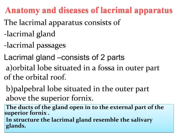
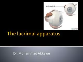
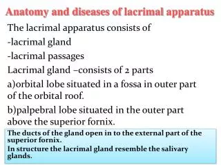
Anatomy and diseases of lacrimal apparatus. The lacrimal apparatus consists of -lacrimal gland -lacrimal passages Lacrimal gland –consists of 2 parts A-orbital lobe situated in a fossa in outer part of the orbital roof. B-palpebral lobe situated in the outer part above the superior fornix.

E N D
Anatomy and diseases of lacrimal apparatus The lacrimal apparatus consists of -lacrimal gland -lacrimal passages Lacrimal gland –consists of 2 parts A-orbital lobe situated in a fossa in outer part of the orbital roof. B-palpebral lobe situated in the outer part above the superior fornix.
Diseases of the lacrimal apparatus • 1-Acute dacryoadenitis = occurs occasionally in general infection –mumps since the structure of the lacrimal gland is similar to the parotid and submandibular salivary glands. The gland becomes enlarged and tender. There is congestion and chemosis of the congunctiva. • Treatment • 1-Analgesic ,hot fomentation • 2-Systemic antibiotics
Tumores of the lacrimal glands= • owing to similarity of structure tumours of the lacrimal gland show a very marked resemblance to those of the partoid.The commonest one is pleomorphic adenocarcinoma or mixed tumour. It is seen in persons above the age of 40.there is proptosis and in addition the eyeball is pushed downwards and inwards.
Sjogrens syndrome or keratocnjunctivitis sicca • It is autoimmune disease seen in women after • menopause and often associated with rheumatoid arthritis. • Clinical features • It is characterised by deficiency of the lacrimal secretion leading to dryness of the eye. • There are chronic irritative symptoms.The cornea shows superficial punctate or a filamentary keratitis. Damage to the epithelium of the conjunctiva and cornea may be demonstrated by staining pink with rose bengal.similar affection of the salivary glands may lead to dryness of mouth and the stomach to achlorhydria
Treatment • Tear substitutes
The ducts of the gland open in to the • external part of the superior fornix • In structure the lacrimal gland resemble • the salivary glands.
Lacrimal passages –consist of • A- lacrimal puncta- one in each lid and situated near the posterior border of the margin 6 mm from The medial canthus. • B- Canaliculi – one in each eyelid-it commences at the punctum and carries tears to the lacrimal sac. • C- Lacrimal sac =lies in the lacrimal fossa formed by the lacrimal bone and frontal process of maxilla.It is covered by lacrimal foscia. • Anterior to the lacrimal foscia is the medial palpebral ligament. • The upper part of the sac is known as the fundus.The lower end narrows as it open into the nasolacrimal duct.
D-Naso lacrimal duct It is 3-4 in • length and • opens into the inferior meatus of the nose. It is directed downwards,slightly outwards and backwards. • The tears which are secreted by the lacrimal glands into the conjunctival sac are drained by the lacrimal passage into the nose.
Watering of the eye • 1-Epiphora –denotes watering due to obstruction of outflow of tears • A- Eversion of the lower punctum in ectropion of lower lid. • B-Occlusion of the lower punctum • congenital • Acquired cicatricial • C- Occlusion of the lower canaliculus due to • a scar, foreign body-eyelash,fungus, • D-Chronic dacryocystitis
2-Lacrimation- reflex – • causes due to sensory stimulation as in • corneal ulcer ,exposure to smoke and irritant gases.
Chronic Dacryocystitis • Etiology = It is usually due to an obstuction in the nasolacrimal duct,followed by infection of the lacrimal sac. • 1-Obstruction of naso lacrimal duct • a-congenital • B-Tubercululosis,syphilis and leprosy –oringinating in surrounding bones or the nose. • C- Stricture of the duct due to chronic atrophic rhinitis. • d-Obstruction of the lower end of the duct due to nasal polypi,hypertrophied inferior turbinate bone,extreme deviation of nasal septum. • E- Maxilla –fructure,maxillary sinusitis, tumours of maxillary antrum. • F= Tumours of the lacrimal sac
2-Infection – • stagnation of the content of lacrimal sac is quickly followed by infection.as a result of contamination by micro organisms from conjuctiva- staph ,strept, and pneumoccocci apurulent inflamation of thr lining of the sac is established
Sings • –it occurs more commonly in women. • Age 40-60 years age group. • 1-There may be pooling of tears near the media canthus. • 2-On pressure over the sac there may be a mucoid or mucopurulent reguritation through the puncta or more rarely it passes down the nose. • 3- There may be a non tender swelling
Complications • 1-chronic conjunctivitis • 2-Acute dacryocystitis may arise. • 3-Lacrimal abscess. • 4-Lacrimal fistula- when the lacrimal abscess rupturees or is drained. • 5-Orbital cellulitis,facial cellulitis and rarely cavernous sinus thrmbosis.
Treatment • 1-In the new born • A=gentamycin drops • b-=Massage • C=Probing of the naso lacrimal duct • In adults • 1=Dacryocystectomy • 2-DCR
Acute dacryocystitis • It occurs usually as an acute exacerbation of chronic dacryocystitis.Rarely it may start spontaneously.The same organisme like staph,pneumococci, which cause chronic dacryocystitis also give rise to acute dacryocystitis.
Symptomradiating frontal to the rgion.s • Symptom • 1-Fever • 2-Sever pain over the lacrimal sac area • radiating to the frontal region
Sings • 1- The skin over the sac becomes red ,swollen,warm an tender • 2 -congestion and chemosis of congunctiva • 2-Redness and swelling rapidly extend to the lid and upper part of cheeks.
Treatment • 1-Analgesics • 2- hot fomentation • 3- A-B