Download
1 / 48
530 likes | 945 Views
Gastrointestinal Intubations. Module 5. Gastrostomy Surgical creation of a gastric fistula through the abdominal wall, necessary in some cases of cicatricial stricture of the esophagus for the purpose of introducing food into the stomach. Percutaneous endoscopic gastrostomy (PEG)

E N D
Gastrointestinal Intubations Module 5
Gastrostomy • Surgical creation of a gastric fistula through the abdominal wall, necessary in some cases of cicatricial stricture of the esophagus for the purpose of introducing food into the stomach. Percutaneous endoscopic gastrostomy (PEG) • A feeding ostomy. PEG tubes are inserted through the esophagus into the stomach with the aid of an endoscope and then pulled through a stab wound made in the abdominal wall. Nasogastric • the nostril to the nasopharynx and to the stomach
Ileus • failure of appropriate forward movement of bowel contents. It may be secondary to either mechanical obstruction of the bowel or a disturbance in neural stimulation (adynamic ileus). Paralytic ileus • paralysis of the gut due to peritoneal contamination by pus (from a perforated appendix) or acid (from a perforated ulcer); • Underlying condition must be treated appropriately • Can also follow spinal cord injuries, pneumonia, and other general conditions. May occur after any abdominal surgery & can be a anticholinergic side effect of certain psychotropic drugs. • Usually temporary, with distention & symptoms of acute obstruction
Gastrointestinal Tubes • Can be inserted via • Nose • Stomach • Many types can be inserted by nurses, but some of the larger, longer or multiple lumen tubes must be inserted by physicians or nurses with advanced training
Improper tube placement can lead to serious complications: • Esophagotracheal fistula • Gastric ulceration • Esophageal or gastric perforation • Aspiration pneumonia
GI tubes • May be inserted for the purpose of • Instilling liquid foods or other substances • Establish a means for suctioning stomach contents to prevent gastric distention, nausea & vomiting • To obtain stomach contents for lab work • To lavage stomach in cases of poisoning or overdoses • Gastric decompression (to remove gas and or fluid)
Enteral Access devices • Access can be achieved via various routes • Nasogastric = inserted thru 1 of nostrils down nasopharnyx to alimentary tract (may also go mouth down but causes gagging++ and discomfort, more often in premies) • Nasoenteric inserted same as Nasogastric but extends down into upper small intestine (used with those with high risk of aspiration) • Gastrostomy, Jejunostomy = for longer term and placed via placement thru abd wall into stomach (gastrostomy) or into jujumen (Jejunostomy)
Nasoenteric tube (NET) is generally indicated for short-term therapy (< 6wks) • More permanent enterostomal devices for nutrition, such as a gastrostomy or jejunostomy tube, is appropriate for therapy expected to last a month to 6 weeks or more.
Types of GI tubes Large Bore used for gastric decompression or analysis, or rarely for short-term feeding • Levin = single lumen tube Size 12 to 18F • Salem = double lumen, larger lumen for draining of gastric contents, smaller allow for inflow of air to prevent a vacuum if tube adheres to stomach wall Small Bore • Nasogastric/feeding tubes – size 6 to 12F and 30-60 inches long Intestinal Tubes for decompression Hg-weighted balloon tips facilitate passing the tube beyond the stomach primarily for intestinal decompression (10 feet long) • Harris, Cantor or Miller Abbot
Smaller diameter • Smaller diameter tubes • Clog easier esp with thick feeds or when meds are tabs that have been crushed • Can also be dislodged by vomiting or coughing • Can become knotted or kinked in GI tract • If not flushed before and after, easily become clogged requiring a change of tube • More difficult to check residual volumes
Tubes made of polyurethane or silicone • Radiopaque so position visible by x-ray • Tubes made of rubber or polyvinyl chloride stiffen with time
Procedures • Inserting a NG tube – Kozier p. 1149 • Removing NG tube – Kozier p. 1152 • Managing GI suction – Kozier p. 872 • Administering tube feed via NG tube – Kozier p. 1156 • Administering feed via gastrostomy or jejunostomy feed – Kozier p. 1158
Purposes of NG tubes • Decompression (remove fluids & gas from stomach) • Prevent or relieve N&V after surgery (by the decompression) • To determine amt of pressure & motor activity in GI tract • Irrigate stomach (lavage) after poisoning • Treat mechanical obstruction • Administer meds & nutrition • Obtain specimens (when pyloric or intestinal obstruction suspected)
NG tubes • Used with • Pts with intact gag & cough reflexes • Pts with adequate gastric emptying • Short term use
Inserting a NG tube • If rubber tube used, place on ice for 5-10 mins to stiffen tube • Make sure guide wire if present is secured in position • Measure distance (NEX) • Use only water soluble lubricant (muko) not Vaseline to lubricate tube • Have pt sit upright, head back • Once tube at throat (gag) have client tilt head forward and swallow • NEVER, NEVER, EVER FORCE A TUBE DOWN
Obtaining the NEX (nose, earlobe and xiphoid) Some tubes may be pre-measured but this may not correlated exactly with the measurement obtained
TO CONFIRM PLACEMENT OF NG • Placement most reliably confirmed by x-ray • Can also aspirate for stomach contents or auscultate air insufflation • Aspirate for stomach contents = attach syringe to tube and apply gentle pressure. If contents return, tube is in place • Air insufflation = attach syringe, place stethoscope over pt’s epigastric region. Inject about 15-20ml of air and listen for “whoosh” • Ask pt to talk = if cannot talk, tube may be coiled in throat or passed through vocal cords
Methods to assess tube position • Test gastric aspirate for pH (1 – 4 – 6 {if pt on meds for gastric acid control}) • Intestine aspirates at pH 6 & > • Resp aspirates at 7 & > • pH readings of 6+ indicate need for x-ray confirmation
Pt with NG tube • Will require • Inspect nose for discharge and irritation • Clean nostril and tube with moist q-tips • Apply water soluble lubricant to nostril if area dry, crusted • Frequent mouth care (q2h) as client will be NPO
NG problem solving • Pain or vomiting after insertion = indicates tube is obstructed or incorrectly placed • Not draining = may be obstructed or needs to repositioned then checked again for placement • If pt displaying signs of distress (gasping, coughing, cyanotic) remove tube immediately
Nurses responsibility • Assess gastric contents • Color • Consistency • Odor • Amount • Irrigate tube before and after meds/feeds • Check GI function by auscultation for bowel sounds
Key Elements in insertion of NG • Minimize discomfort by positioning pt appropriately, lubricating tip and informing pt what to expect • NEVER force the tube against resistance • Confirm placement of the tube • Secure the tube to the pt’s nose or nose and cheek
NG’s & suction, decompression Kozier on p. 872 • Suction can be ordered as • Continuous (can be set anywhere from 60-120 mm Hg) • Intermittent (suction for 15-60 secs then interval with no suction) usually at low setting And • Low(pressure between 80-100 mm Hg) • High (pressure between 100–120 mm Hg
Can be connected to • Wall suction • Portable suction • By gravity • Before initiating, 1st confirm tube in place • Intermittent – usually with single lumen • Continuous – usually with double lumen (Salem)
NG’s & suction, decompression… • Low continuous suction or high intermittent pressure = Salem sump tube (ie has double lumen. • Larger lumen for draining of gastric contents, smaller allow for inflow of air to prevent a vacuum if tube adheres to stomach wall • If Levin tube used, low intermittent suction recommended
Key Elements to Remove NG • Turn off suction, disconnect tube from suction • Clamp tubing before removal • Wear gloves • Withdraw tube while pt holds his breath
Part B: Enteral Feeding & Meds • Oral feeding is preferred & most effective method but there are alternates for po nutrition including: • Enteral (thru GI system) and can be via • NG tubes • Gastrostomy or jejunostomy tubes • Parenteral (IV) • Alternates may deliver total or supplemental nutrition over short or long term
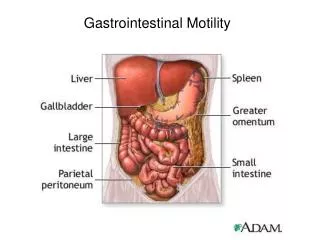
Common enteral feeding placement locations From Lewis on p. 1054
NG tubes for feeds • Until recently, large bore rubber or plastic feeding tubes were used for NG feeds. Problems with tubes (irritation, pharnygitis etc) led to development of more pliable, small bore feeding tubes • Now the are 8-12 Fr (36-43”long) • Have weights to assist introduction but also keep in place • Problem with smaller tubes is the difficulty to verify placement (difficult to aspirate & insufflating with air unreliable as get false positives – can sometimes hear the gurgle even if the tube is in the esophagus or lung)
Enteral Feeding & Meds • Short term – less than 6 wks • A NG or nasointestinal tube appropriate • Advantages of NG feeds: • Allows stomach to be used as natural reservoir (so regulates amt of food/liquid released into small intestine) • Presence of gastric juice may decrease risk of infection • Disadvantages of NG feeds • Potential aspiration into lungs • Uncomfortable • Pt’s with dysfunctional gag reflux or those unable to be in Fowler’s position not candidates
Enteral Feeding & Meds • Nasointestinal Tubes – inserted via nostril to upper portion of small intestine • Advantage: minimal risk for aspiration • Disadvantage: may develop dumping syndrome as food direct in ‘bolus’ into intestine (as pyloric valve in stomach cannot slow or regulate transit of food into intestine) • Volume of feed results in • Distention of intestine • Hypoglycemic reaction • Results in gas, bloating, crampy pain, weakness & dizziness
Enteral Feeding & Meds • Long Term Support accomplished by creating a an opening into: • Gastrostomy (opening into stomach) • Jejunostomy (opening into jejunum) • Methods of feeding includes: • PEG (Percutaneous endoscopic gastrostomy) • Surgical or laproscopically placed gastrostomy tube
More & more PEG’s are popular as can be safely inserted & removed at bedside or in OPD To insert requires (by a physician) • local anesthesia, • passage of endoscope into stomach, • a small incision or stab wound thru the skin of the abdomen • pushing a cannula thru the incision • insertion of guide wire thru cannula • introduction & placement of PEG
Before feeds with PEG’s or NG • NG’s or NI’s placement should be confirmed: • After insertion • Before beginning meds or feeds • At regular interval during a continuous feed • Esp important with the smaller bore tubes (ie. 6-12FR)
With NG’s and New PEG’s anticipate: • Measuring of residual before each feed • Measuring residual done to evaluate absorption of last feed (ie. Is there undigested formula from a previous feed) If residual is more than last infusion or 150 ml, hold feeding for 1 hour and recheck. For continuous feeds, check residual q4-6 hours
Assess bowel sounds prior to each feeding or for continuous feeds, q4-8h • Monitor for abdominal distention (would indicate intolerance to previous feed) • Monitor for diarrhea, constipation or flatulence (lack of bulk may cause constipation. Hypertonic or concentration of formula may cause diarrhea or flatulence)
Nursing care • Elevate head at least 30 degrees during feed and 1 hr following • Tube must be flushed before and after feeds, med admin or aspiration for patency • Delivery sets must be changed q 12-24hrs or according to policy
Nursing care • Opened cans must be stored according to manufacturer’s directions • Clean skin and stoma area at least daily, should be qshift • Mouth care q2-4 hours