Download
1 / 59
640 likes | 1.2k Views
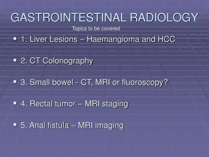
GASTROINTESTINAL RADIOLOGY. Topics to be covered. 1. Liver Lesions – Haemangioma and HCC 2. CT Colonography 3. Small bowel - CT, MRI or fluoroscopy? 4. Rectal tumor – MRI staging 5. Anal fistula – MRI imaging. Liver – Haemangioma (US). Atypical. Liver Haemangioma CT A) Pre-contrast.

E N D
GASTROINTESTINAL RADIOLOGY Topics to be covered • 1. Liver Lesions – Haemangioma and HCC • 2. CT Colonography • 3. Small bowel - CT, MRI or fluoroscopy? • 4. Rectal tumor – MRI staging • 5. Anal fistula – MRI imaging
Liver – Haemangioma (US) Atypical
D) Delayed phase CT – we will not do delayed phase unless haemangioma suspected. Please specify “? haemangioma” on request form.
Haemangioma Summary • Common- often incidental • US – Echogenic -no halo. No colour flow. Aytpical – hypo-echoic in fatty liver - mixed echotexture • CT – C- low density C+ peripheral vessels (uneven) C+ PV /delay progressive fill-in Small haemangioma fill in immediately and cannot be distinguished from metastates. • MRI features similar to CT post Gadolinium
HCC Summary • US - usually heterogeneous Usually HepB +ve with raised alpha FP • CT – C- low density C+A – central early contrast (high flow rate) C+PV – washout cf with liver – may have a capsule • MR – intracellular fat on T1 out of phase - similar perfusion characteristics to CT
MRI IMAGES of LIVER • Look at CSF first to tell if T1 or T2 • T1-in/out. • T1 are grey. Fluid is dark. Black outline • T2-incl HASTE. • More definition. Fluid is bright. • Gadolinium – always with T1
CT COLONOGRAPHY Dissection Strip, anus to caecum Endoluminal (for fun only) Orientation Overview 800/40 window Axial to loops
Advantages / disadvantages • Sensitivity and specificity is of the order of 90 % for 10 mm polyps. • Easy, quick and well tolerated. • Beats barium enema hands down. • Safer than optical colonoscopy • Approx. half the price of optical colonoscopy • No intervention possible as in optical Cy • At present for “Ba enema” indications, but is likely to be used for screening in future. • Radiology manpower training required. • Radiation dose equivalent to Ba Enema
CTC vs Optical Colonoscopy Consider “Is intervention likely to be needed?” – (cf MRCP vs ERCP) • CTC for average risk and Fam Hx pts. • > 50 yrs (radiation) • Contraindicated if inflammatory bowel or on steroids (risk of perforation as inflation is done “blind” as opposed to Ba enema). • Optical Colonoscopy – if biopsy or polypectomy prob needed • All polyposis syndromes • High risk • Inflammatory Bowel Disease
Overview of CT colonography? • Process Currently Future • CLEANSE -Tagging -Subtraction • DISTEND -Air -CO2 • COMPUTE -Workstation -new programs • VIEW -Time - CAD • REPORT -Issues
Prep and tagging Slide courtesy Dr Helen Moore
Longer tube and patient can apply air themselves Slide courtesy Dr Helen Moore
Incomplete air column -Excess fluid Supine Prone Can rotate image volume to view as a Ba enema in 3D
Ileo-caecal valve Caecal pole Arrow points To caecum Residual tagging
Dirty Caecum- not fully open on supine or prone views 54 yr Recomm optical colonoscopy
Radiation • Barium enema 6 – 8 mSv • CTC estimate of 7.6 mSv with low mAs. Increased noise, but high resolution improves definition of small polyps • Thin slice, limit tube current • Background radiation is 2.4 MSv/year The worldwide average background dose for a human being is about 2.4 millisievert (mSv) per year.[1] This exposure is mostly from cosmic radiation and natural isotopes in the Earth. This is far greater than human-caused background radiation exposure, which in the year 2000 amounted to an average of about 0.01 mSv per year from historical nuclear weapons testing, nuclear power accidents and nuclear industry operation combined,[2] and is greater than the average exposure from medical tests, which ranges from 0.04 to 1 mSv per year. Source Wikipedia.
Small Bowel Imaging • < 35 yrs – MRI for radiation reasons • However if pre-surgical workup–fluoroscopy • CT Enteroclysis – only difference from CT is negative contrast in bowel. No advantage to do if recent normal CT. • MR Small bowel – breath-hold sequences, dynamic change between sequences. Good soft tissue differentiation. +/- Gadolinium
Normal Fluoroscopic Enteroclysis Jejunal intubation Low density barium Pumped in to distend Intubation 10 min Study 20 min
Follow-throughtime-consumingflocculationStrictures may be hiddenIs superseded by other tests
CT Enteroclysis Histo- GIST Tumor shows up against negative contrast in bowel. Positive contrast could hide it
CT ENTEROCLYSIS Volumen oral contrast for 45 min pre scan IV Maxolon IV contrast on table CT to include anal canal and with sagittal.
CT ENTEROCLYSIS Jejunum often thick-walled Can evaluate bowel wall due to negative contrast in lumen and IV contrast in wall. Evaluates stomach well also Plus standard CT Reserved for older patients due to radiation dose
MRI Small Bowel • Oral Volumen 30 – 45 min prior (or Ioscan) • +/- IM Buscopan for peristaltic movement • Good for Crohns patients with multiple studies and large radiation dose over time. • Coronal TRUFI • Coronal TRUFI fat saturation • Coronal HASTE • Axial HASTE • Coronal T1
Cutaneous fistula Post Gadolinium T1 fat sat
Normal FAT SATURATION