Download
1 / 33
380 likes | 564 Views
Crystal Structure of Argonaute and Its Implications for RISC Slicer Activity. Ji-Joon Song, Stephanie K. Smith, Gregory J. Hannon, Leemor Joshua-Tor. Pamela Lussier Biochemistry 4000/5000. RNA interference. http://www.nature.com/focus/rnai/animations/animation/animation.htm.

E N D
Crystal Structure of Argonaute and Its Implications for RISC Slicer Activity Ji-Joon Song, Stephanie K. Smith, Gregory J. Hannon, Leemor Joshua-Tor Pamela Lussier Biochemistry 4000/5000
RNA interference http://www.nature.com/focus/rnai/animations/animation/animation.htm
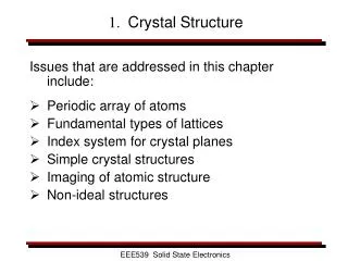
RNA interference (RNAi) • Triggered by the presence of dsRNA • RNase III family enzyme Dicer • initiates silencing by releasing siRNA ~ 20 base duplexes with two-nucleotide 3’ overhangs • siRNA guide substrate selection by effector complexes called RISC
RISC (RNA induced Silencing Complex) • Contain single stranded versions of siRNA as well as additional protein components • One of which is a member of the Argonaute family of proteins • RISC recognizes and destroys target mRNAs by cleavage in region homologous to siRNA.
Argonaute Protein • Defined by presence of PAZ (Piwi Argonaute Zwille) and PIWI (named for protein piwi) domains • PAZ domain of Argonaute interacts directly with small RNA in RISC • Forms a oligonucleotide/oligosaccharide binding (OB) fold containing a central cleft lined with conserved aromatic residues that bind specifically to single stranded 3’ ends
In RISC, the Argonaute PAZ domain would hold the 3’ end of the single stranded siRNA • Possibly orients recognition and cleavage of mRNA substrates. • Nuclease Responsible for cleavage (Slicer) has escaped identification.
Objective • Deepen understanding of the role of Argonaute protein in RNAi • To conduct structural studies of a full length Argonaute protein from Pyrococcus furiosus.
Hanging Drop Method • Initial crystals were grown by vapor diffusion using hanging-drop method in presence of small amounts of organic solvent
Quality of crystals improved by microseeding Control nucleation as it consists of introducing crystal nuclei in the equilibrated metastable protein solution, where seeds might grow
Structural Determination • Multiple Anomalous Dispersion (MAD) • Used selenomethionine substituted protein crystal. • Structure of full-length Argonaute (PfAgo) determined to 2.25 Angstroms.
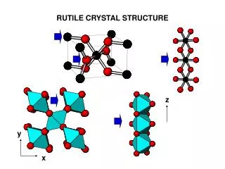
Overall architecture • N-terminal, middle, and PIWI domains form a crescent-shaped base • N-terminal domain forms a “stalk” that holds PAZ domain above the crescent and an interdomain connector cradles the four domains of the molecule. • Forms a groove at the center of the crescent and the PAZ domain closes off the top of this groove.
PAZ Domain Red = PfAgo Gray = hAgo1 Dotted lines in figure represent disordered regions
Sequence Alignment of PAZ domains of PfAgo, hAgo1 and DmAgo2 Primary sequence comparisons failed to reveal PAZ domain despite close structural similarities Purple = invariant residues Blue = conserved residues
PAZ domain • Conserved aromatic residues that bind the two-nucleotide 3’ overhang of an siRNA are present in PfAgo. • Side chains occupy similar positions in space, but they are anchored to peptide backbone in different locations. Green = hAgo1 residues
So…where does the 3’ overhang of the siRNA bind??? Right Here!!!
PAZ Domain Comparison • L263 and I261 assume role of L337 and T335 in hAgo1, which anchor sugar ring of terminal residue through vand der Waals interactions. • W213 assume role of F292 in hAgo1, which stacks against the terminal nucleotide. • R220 is positioned similarly to K313 that contacts the penultimate nucleotide. • Reasoned that the PAZ domain in PfAgo binds RNA 3’ ends, as do PAZ domains of fly and human Argonautes.
PIWI is an RNase H Domain The domains are topologically identical Five-stranded mixed β sheet surrounded by helices
PIWI Domain • 3 highly conserved catalytic carboxylates • One is located in β1, and one is located at C terminus of fourth strand β4 • The third carboxylate varies • Only requirement is a reasonable spatial position at the active site.
Active Site Rotated 180° Two aspartate residues in PIWI were located at same positions as the invariant carboxylates – D558 on first β strand, and D628 on the end of the fourth strand. E635 is in close proximity to the two aspartates and suggests that this glutamate serves as the third active-site residue.
Active site is positioned in a cleft in the middle of the crescent in the groove below the PAZ domain. Here on overall structure
Ago is Slicer • Argonaute is the enzyme in RISC that cleaves the mRNA. • RNase H enzymes cleave ssRNA “guided” by DNA strand in RNA/DNA hybrid. • Argonautes might do RNA cleavage guided by the siRNA strand in a dsRNA substrate. • Depends on Mg+2, like other RNase H enzymes.
Distinct groove throughout the protein, which has a claw shape and bends between the PAZ and N-terminal domains. Electrostatics show inner groove is lined with positive charges – suitable for interaction with negatively charged phosphate backbone. Blue = positively charged Approx location of active site marked by a yellow asterisk
Possible substrate binding • Superimposed PAZ domains from PfAgo and hAgo1 and examined position of RNA in hAgo1 complex with respect to PfAgo. • siRNA guide interacts with PAZ cleft
Model for siRNA-guided mRNA cleavage siRNA binds with its 3’ end in the PAZ cleft and the 5’ is predicted to bind near the other end of the cleft. The mRNA comes in between the N-terminal and PAZ domains and out between the PAZ and middle domain. The active site in the PIWI domain cleaves the mRNA opposite the middle of the siRNA guide.
From studies of other RNase H enzymes, expected that Argonaute senses the minor groove width of the dsRNA, which differs from that of dsDNA. • Fits with RISC’s inability to cut DNA substrates. • Opening of the claw might assist binding of mRNA – hinge region exists in interdomain connector at residues 317-320.
Added Support • In mammalian system, performed mutational analysis on hAgo2 • Conserved active site aspartates were altered = loss of nuclease activity but retained siRNA binding.
Remaining Questions? • Other determinants beyond catalytic triad of PIWI domain that determine activity toward RNA substrates, such as conformational differences. • Interactions with other factors may be needed to create fully active Slicer
Identification of catalytic center of RISC awaited a drive towards understanding RNAi at a structural level. • A full understanding of the underlying mechanism of RNAi will need to be derived from a combination of biochemical and structural studies of RISC.